
Figure 5: Increases of Tyrobp and Apoe mRNAs in microglia recruited to a site of stab injury are Trem2-independent.
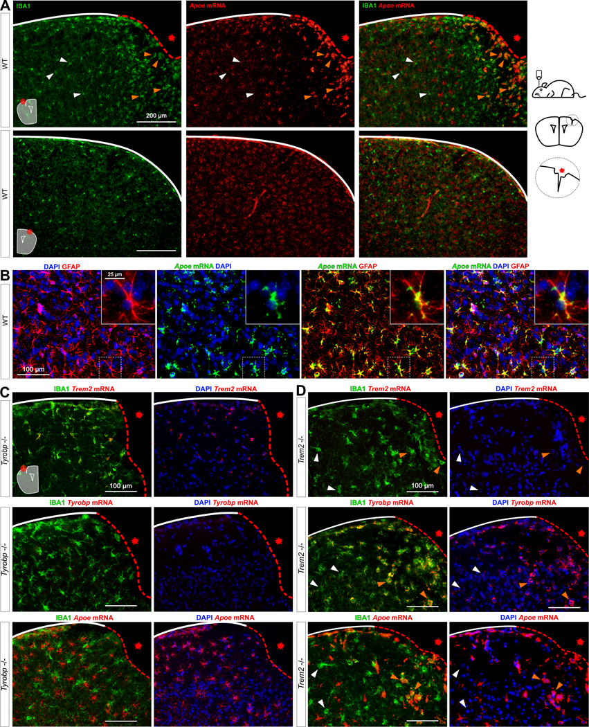
(A) Stab-injured WT mice were sacrificed 3 days after injury and dual RNA fluorescent in situ hybridization and immunohistochemistry for Apoe mRNA (red) and anti-IBA1 (green) respectively was performed. The injured ipsilateral area (red dotted line) is shown on the top row and the uninjured contralateral area is shown on the bottom row. Scale bar = 200 μm. (B) Dual RNA fluorescent in situ hybridization and immunohistochemistry for Apoe (green) and GFAP (red) in non-injured WT mice. (C-D) The same stab injury protocol was utilized in Tyrobp−/− (C) and Trem2−/− (D) mice. Anti-IBA1 staining and DAPI staining are shown in green and blue, respectively. Top row: Trem2 mRNA (red); middle row: Tyrobp mRNA (red); bottom row: Apoe mRNA (red). Mice were 4 months of age, and slice thickness = 10 μm. The red asterisk indicates the injured side. White and orange arrows indicate examples of non-recruited and recruited microglia, respectively.