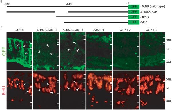
Figure 1. A 109 bp region of the α1T promoter is required for transgene expression in dedifferentiating Müller glia.
(a) Schematic representation of α1T promoter constructs. The bars represent promoter sequence and the numbers indicate relative position from the start codon. −1696 is the wild-type promoter described previously15,16. Δ-1046-846 has been described17. The −1016 promoter directs transgene expression in Müller glia1. The −907 promoter lacks 789 bp of upstream sequence. (b) Transgenic fish received retinal injuries on day 0 and were given a 4 hr pulse of BrdU at 4 days post injury. Transgenic fish which carry the required DNA element express GFP in BrdU labeled Müller glia (−1016 panel, arrows), while transgenic fish lacking the element do not (Δ-1046-846 and −907 panels). Two independent lines of Δ-1046-846 and three independent lines of −907 transgenic fish all display a lack of GFP expression in BrdU labeled cells (arrowheads in Δ-1046-846 panels). The images for −1016 and Δ-1046-846 are from the same sections. Because the −907 transgenic fish display very weak GFP expression in general, we used serial sections to obtain the −907 images. ONL, outer nuclear layer; INL, inner nuclear layer; GCL, ganglion cell layer.

