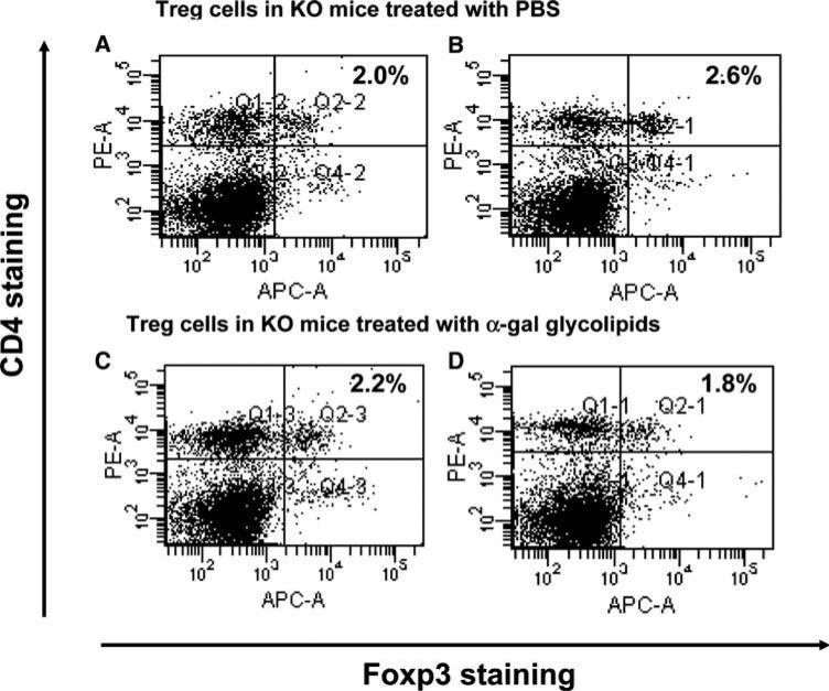
Fig. 6.
Immunostaining of Treg cells from spleens of mice with tumors injected with PBS (a, b), or injected with α-gal glycolipids (c, d). Spleen lymphocytes were obtained 2 weeks after second injection and subjected to Foxp3 and CD4 double staining. The Treg cells are located in the upper right quadrant. Presented data are of two representatives out of five mice in each group

