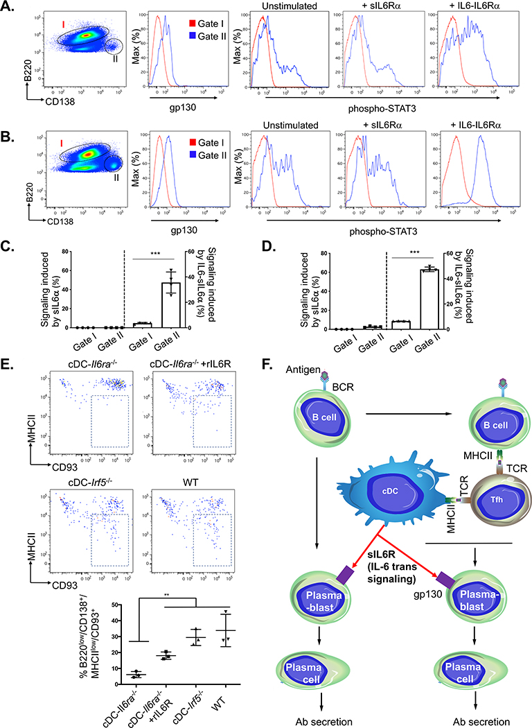
Figure 3. cDC-derived sIL-6R gates antibody responses by controlling plasma cell differentiation.
(A) Surface expression of gp130 on B cells (B220+/CD3−, gate I) versus plasmablasts (CD3−/B220lo/CD138+, gate II) isolated from spleen of WT mice. The splenocytes were either left unstimulated or exposed to sIL-6Rα or IL6-sIL-6Rα (hyper-IL-6).
(B) Same as in (A), except that animals were first immunized with lipid A adjuvant and spleen was harvested 3 days later.
(C) Quantification of the data in (A). Signaling induced within gate I and gate II was measured as the percentage of STAT3-activated cells (sIL-6Rα-stimulated minus unstimulated and IL-6-sIL-6Rα-stimulated minus unstimulated; mean ± SD, n = 4 animals per group, ***p < 0.001, Student’s t test).
(D) Quantification of the data in (B) (as in C).
(E) Differentiation from plasmablasts (CD3−/B220lo/CD138+/MHCII+) to plasma cells (CD3-/B220lo/CD138+/MHC class IIlo/CD93+) was monitored within each genotype 1 week post-immunization with lipid A adjuvant, and cDC-IL6ra−/− mice were also co-injected with recombinant sIL-6R (as in Figure 2E). The proportion of plasmablasts differentiating into plasma cells (indicated by a box in flow plots) was then quantified below (mean ± SD, n = 3 animals per group; **p < 0.005, ANOVA with Tukey’s test).
(F) Model for T cell-independent, cDC-dependent control over antibody responses.

