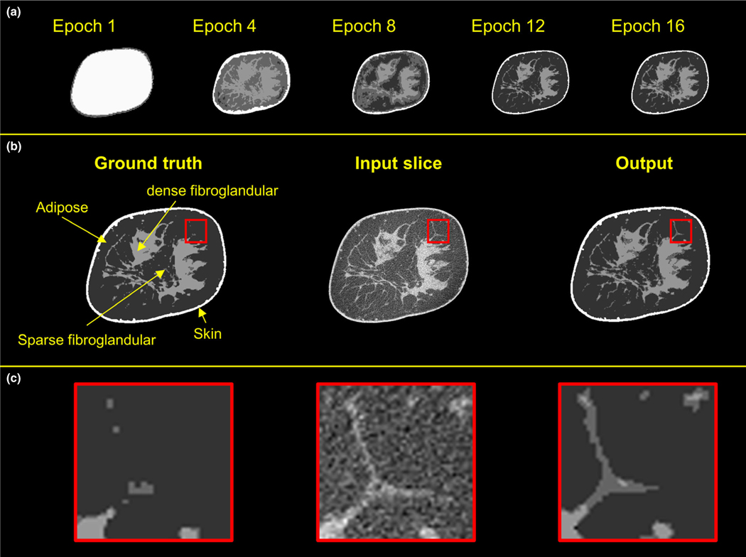
FIG. 4.
Examples of the progression of the network training. Top row shows the output of the network during training. The input coronal image, input segmented image, and the output of the trained network are shown in the middle row. The bottom row displays the zoomed-in views of the regions of interest.

