Figure 2.
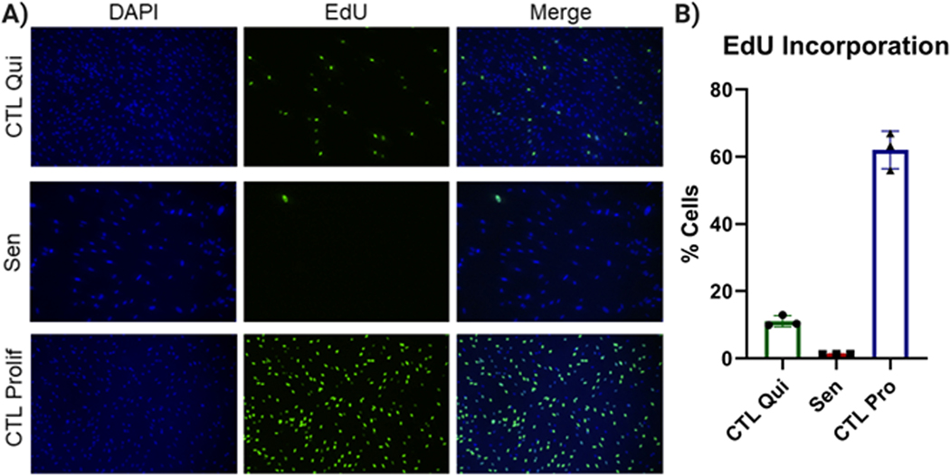
EdU incorporation. A) Representative images of quiescent control (left), IR-induced senescent (center), and proliferating control IMR-90 cells (right). DAPI staining is shown at the top, EdU staining at the bottom. B) Quantification of EdU-positive cells for quiescent control, IR-induced senescent, and proliferating control cells. Data shown are means of 3 replicates ± SD.

