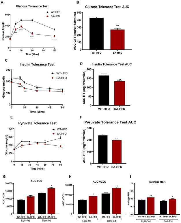
Figure 4. SA knock-in mouse model has improved metabolic capacity under conditions of diet induced obesity.
(A) Glucose tolerance test excursion curve and (B) glucose tolerance test area under curve after 6 weeks of HFD. Data are presented as means ± SEM (n=5/group; **p<0.01, ***p<0.001, ****p<0.0001 vs WT-HFD). (C) Insulin tolerance test excursion curve and (D) insulin tolerance test area under curve after 6 weeks of HFD. Data are presented as means ± SEM (n=5/group; **p<0.01, ***p<0.001, ****p<0.0001 vs WT-HFD). (E) Pyruvate tolerance test excursion curve and (F) pyruvate tolerance test area under curve after 6 weeks of HFD. Data are presented as means ± SEM (WT-HFD n=5, SA-HFD n=9; *p<0.05, **p<0.01, ****p<0.0001 vs WT-HFD). (G) Volume of O2 consumption (H) volume of CO2 production; (I), and respiratory exchange ratio of mice was measured and calculated using CLAMS monitoring system as described previously after 12 weeks of HFD 29. Data are presented as means ± SEM (WT-HFD n=5, SA-HFD n=4; *p<0.05, **p<0.01 vs WT-HFD).

