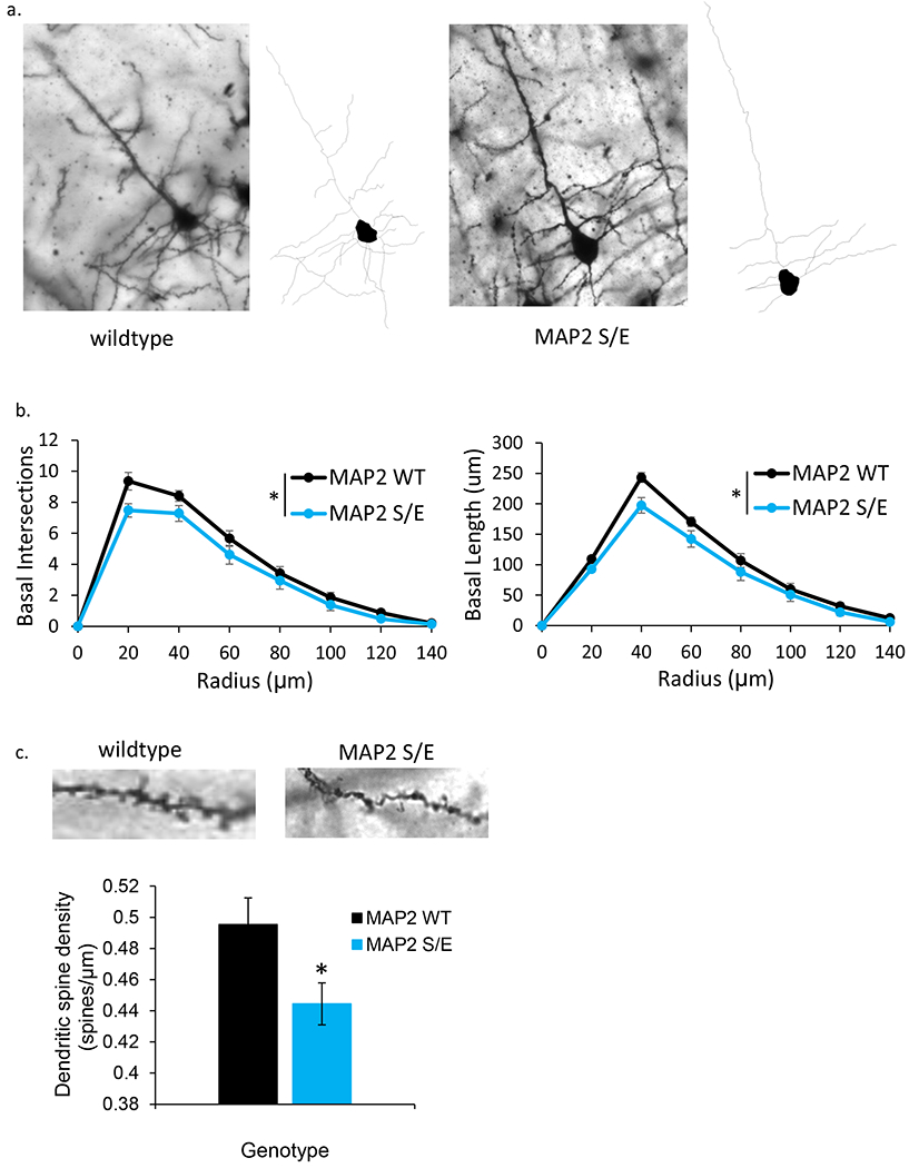
Fig 4. S1782E +/− mice demonstrate reduced basilar arbor branching and length.

(a) Representative single plane Golgi images and corresponding reconstructions of L3 PCs generated from wildtype (left) and S1782E +/− (right) mice. (b) S1782E +/− mice show reduced branching and length in basilar arbors compared to wildtype. (c) Representative single plane Golgi images of a segment of basilar dendrite from a wildtype (left) and S1782E +/− mouse (right). Quantitiatively, S1782 +/− mice show a reduction in spine density along basilar branches compared to wildtype. Sholl data shown are averaged across N=10 animals/genotype (animal values generated by averaging 5 neurons/animal) ± SEM. Spine data shown are generated from the average value per animal across N=10 animals/genotype (animal values generated from 3–5 neurons/animal) ± SEM. *p < 0.05
