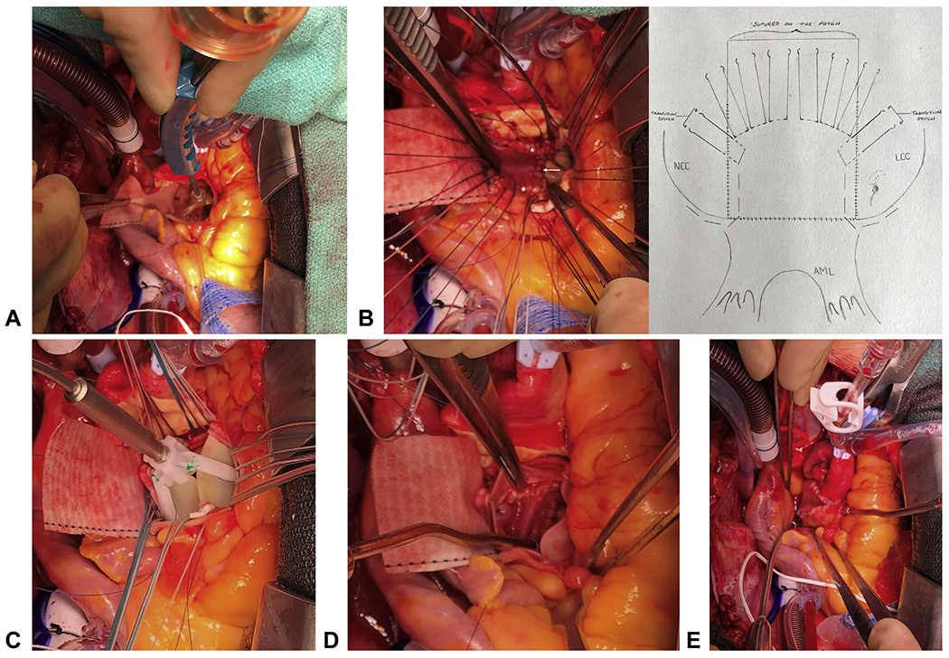
FIGURE 3.

A, The valve position was marked on the Dacron patch to guide the placement of valve sutures with the 2-size larger sizer touching 3 nadirs of the aortic annulus of 3 aortic cusps. B, 2-0 ETHIBOND sutures were placed along the aortic annulus in a non-everting fashion and from outside in on the patch (left panel, white arrow: mitral annulus; white stars: aortic annulus sewn to the patch). The transitional stitches were placed with one arm of the suture through the aortic annulus (non-everting: from ventricle to aorta), and the other arm through the patch (one bite: inside-outside-inside) (rightpanel: illustration). C, The upsized bioprosthetic valve was seated right above the aortic annulus and the Dacron patch with one strut facing the left-right commissure post to position the coronary ostia above the nadir of left and right coronary cusps of the bioprosthesis. D, The left ventricular outflow tract was wide open underneath the upsized bioprosthesis (view from inside of the valve). E, The partial transverse aortotomy was closed incorporating the Dacron patch.
