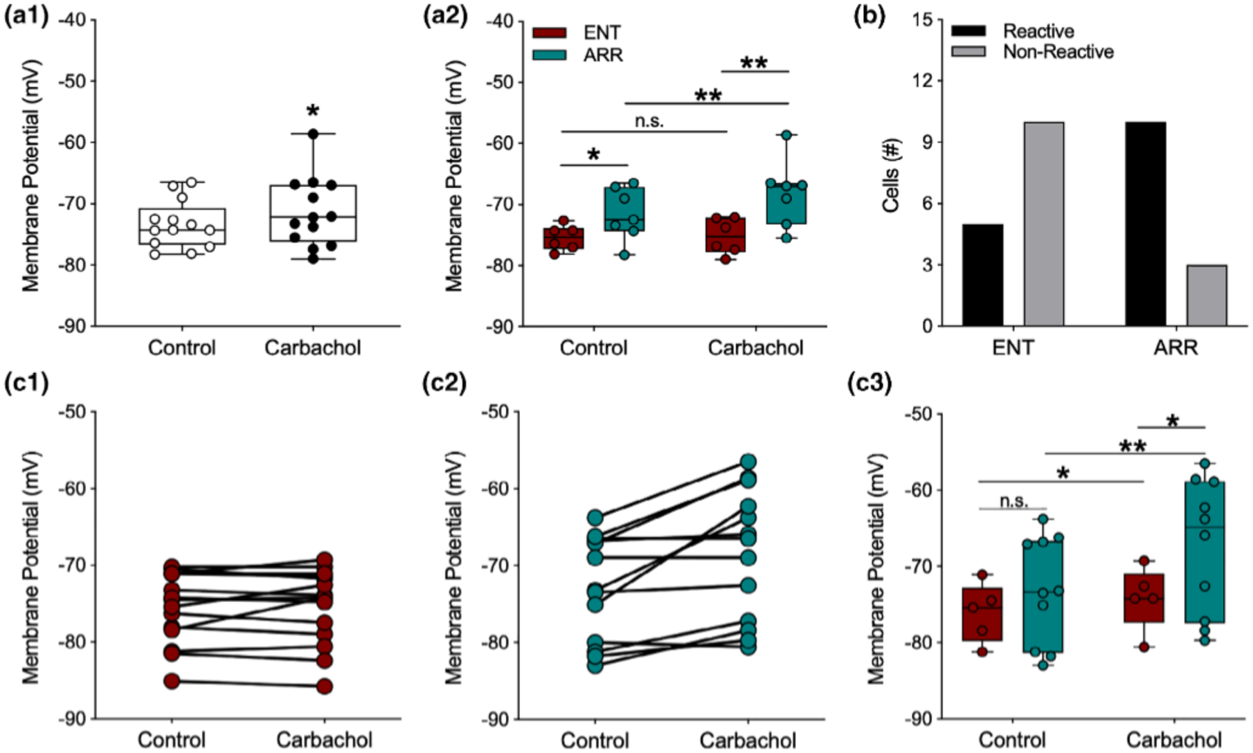
FIGURE 5.

Membrane potentials of dentate granule cells were more depolarized by carbachol in arrhythmic animals. For data points in panel (a2) potentials were averaged across cells within individual animals so that each point is the mean value for a single animal; (a1, b), and (c1–c2) all represent individual cells. For panel (c3), only cells that depolarized under carbachol were analyzed. Carbachol (10 mM), caused significant but modest depolarization of the resting membrane potential (A1; n = 13). However, when data are separated by rhythm status, significant average depolarization only occurred in cells from ARR animals (a2; ENT, n = 6; ARR, n = 7). The resting potential of most ENT cells were not reactive to carbachol (b), while most cells in slices from ARR animals were reactive (b; Fisher’s exact test, p = .008). Membrane potentials of all individual neurons (c1, ENT, n = 15; c2, ARR, n = 13). Membrane potentials only of individual neurons that reacted (depolarized) in response to carbachol (c3). Box plot parameters as in Figure 4. *p < .05, **p < .01
