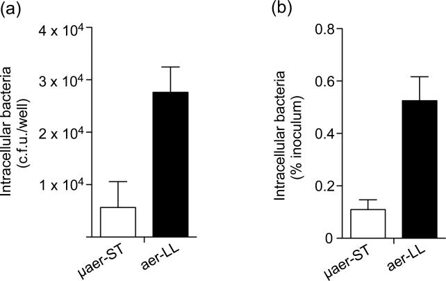
Figure 3. Invasion of epithelial cells by SPI1-induced bacteria.
To directly compare the invasiveness of aer-LL and μaer-ST S. Typhimurium, HeLa cells were infected for 10 min at 37 °C (m.o.i. = 50-60). At 1 h p.i. cells were lysed and intracellular bacteria enumerated by plating. Shown are the means ± SD from three independent experiments. (a) Invasion expressed as c.f.u. (b) Invasion expressed as % of inoculum. In both cases P<0.001, Student's t-test.

