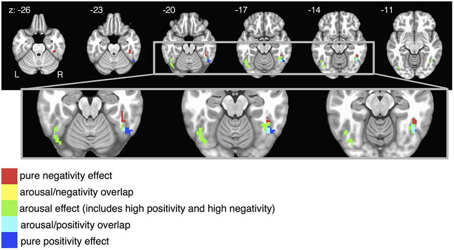
Figure 5.
Fusiform gyrus results from Figure 4A and 4B are combined and focused on in this figure. In the right fusiform, valence is represented along an anatomical gradient. Purely negative-sensitive voxels are more anterior, purely positive-sensitive voxels are more posterior, and arousal-sensitive voxels (which show increased activity to positivity and/or negativity compared to affectively neutral stimuli) are anatomically interposed between the pure valence regions. Combining the results from both theoretical models Valence Arousal and Positivity Negativity are necessary to see this pattern, which would have been overlooked had either model alone had been selected a priori. Colors represent affective condition, not beta weights: all beta weights are positive and survived a reasonably conservative statistical threshold that set the false discovery rate to 0.05.

