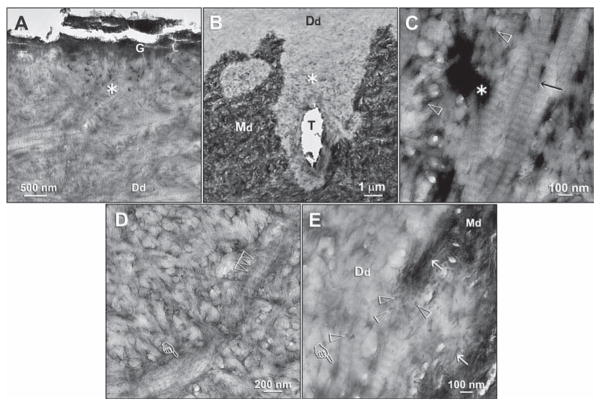
Figure 2.
Ultrastructural characterization of the electron-dense-grains (Ed)-containing zones in the demineralized dentin layers derived from specimens that had been immersed in the remineralization media. (A) A specimen that had been immersed in a PVPA-containing SBF for 4 mos. The electron-dense grainy materials were not exclusively found along the base of the demineralized dentin layer. A higher-magnification view of the junction between the fractured GIC (G) and the underlying demineralized dentin surface revealed an Ed-containing superficial region (asterisk). Although no additional staining was used, excellent banding characteristics of the demineralized dentin collagen matrix (Dd) could be observed. (B) An Ed-containing zone (asterisk) at the base of the demineralized dentin layer. The specimen had been immersed in a polyacrylic-acid- and PVPA-containing SBF for 4 mos. T: dentinal tubule. (C) A high-magnification view of the Ed-containing regions depicted by the “asterisk” in Figs. 2A and 2B revealed a strong negative staining effect, in this bright-field micrograph, that was created in the absence of the use of an extrinsic negative stain. Banding could be identified within the collagen fibrils (arrow) even in the absence of extrinsic staining. No mineral crystallites could be identified around or within these fibrils. Open arrowheads: interfibrillar spaces between the collagen fibrils. (D) A high-magnification view of the area labeled with “Dd” in Fig. 2A. Dark interfibrillar spaces were present despite the absence of the larger, dark terminal tubular channels seen in Fig. 2C. Apart from banding of the collagen fibrils (multiple open arrowheads), the rope-like subfibrillar architecture of the collagen fibril (pointer) was highlighted by the negative staining. No intrafibrillar or interfibrillar crystallites could be identified in these fibrils. (E) A high-magnification view of the junction between the Ed-containing zone and the mineralized dentin base in Fig. 2B. Remnants of apatite platelets (open arrowheads) and needles (arrows) were present along the demineralization front. The Ed within the interfibrillar spaces and terminal tubular branches (pointer) appeared less electron-dense, due to the auto-contrast function of the microscope in the presence of the highly mineralized dentin base along the right side of the micrograph. The absence of apatite remineralization in the negatively stained collagen fibrils and the adjacent interfibrillar spaces is clearly evident. Generic abbreviations: Dd, demineralized dentin; Md, mineralized dentin.

