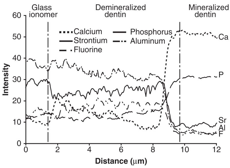
Figure 3.
TEM-EDS line scans of the distribution of calcium (Ca), phosphorus (P), aluminum (Al), strontium (Sr), and fluorine (F) across a representative GIC-dentin interface in a specimen that had been immersed in the SBF devoid of biomimetic analogs for 4 mos. The white dotted line in Fig. 1B represents the generic location upon which elemental analyses were performed across the GIC-dentin interfaces. The changes in elemental profiles may be divided into 3 distinct zones, beginning at the left: (A) In the GIC restoration, strontium, aluminum, and fluorine intensity levels were higher than those in the sound dentin; (B) within the demineralized dentin layer, the 2 GIC-specific elements (strontium, aluminum) were similar to and slightly lower than their corresponding levels in the GIC proper, while, conversely, calcium and phosphate intensities were low within this demineralized layer; and (C) within the mineralized dentin base, the intensity levels of the 2 GIC-specific elements and fluorine decreased to their lowest levels, while those of calcium and phosphorus increased to their maximum levels.

