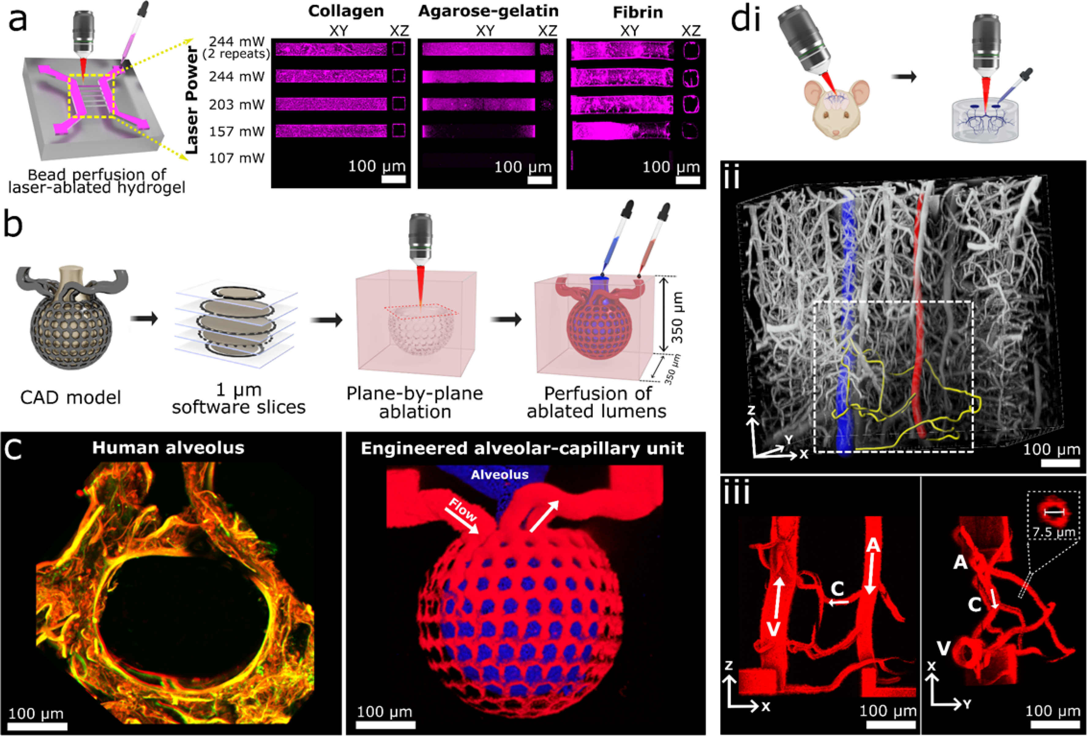
Figure 1: Multiphoton-guided creation of organ-specific vascular structures at anatomic scale.

a, Hydrogel photoablation. Images shown following perfusion of beads (magenta). b, Schematic of ablation and perfusion process. c, Two-photon images of a human alveolus (left), and an engineered alveolar-capillary unit after bead perfusion of capillary (red) and alveolar (blue) compartments (right). d, Recreation of mouse brain microvasculature. i. Schematic of using in vivo imaging data as a mask for ablation. ii: 3D microvascular image traced to isolate an arteriole (red), venule (blue), and capillaries (yellow). iii: Projection images of a replica microvascular unit ablated into collagen and perfused with beads (red). Orthogonal projections and capillary cross-section shown. Arrows delineate flow from arteriole (A), to capillaries (C), and venule (V).
