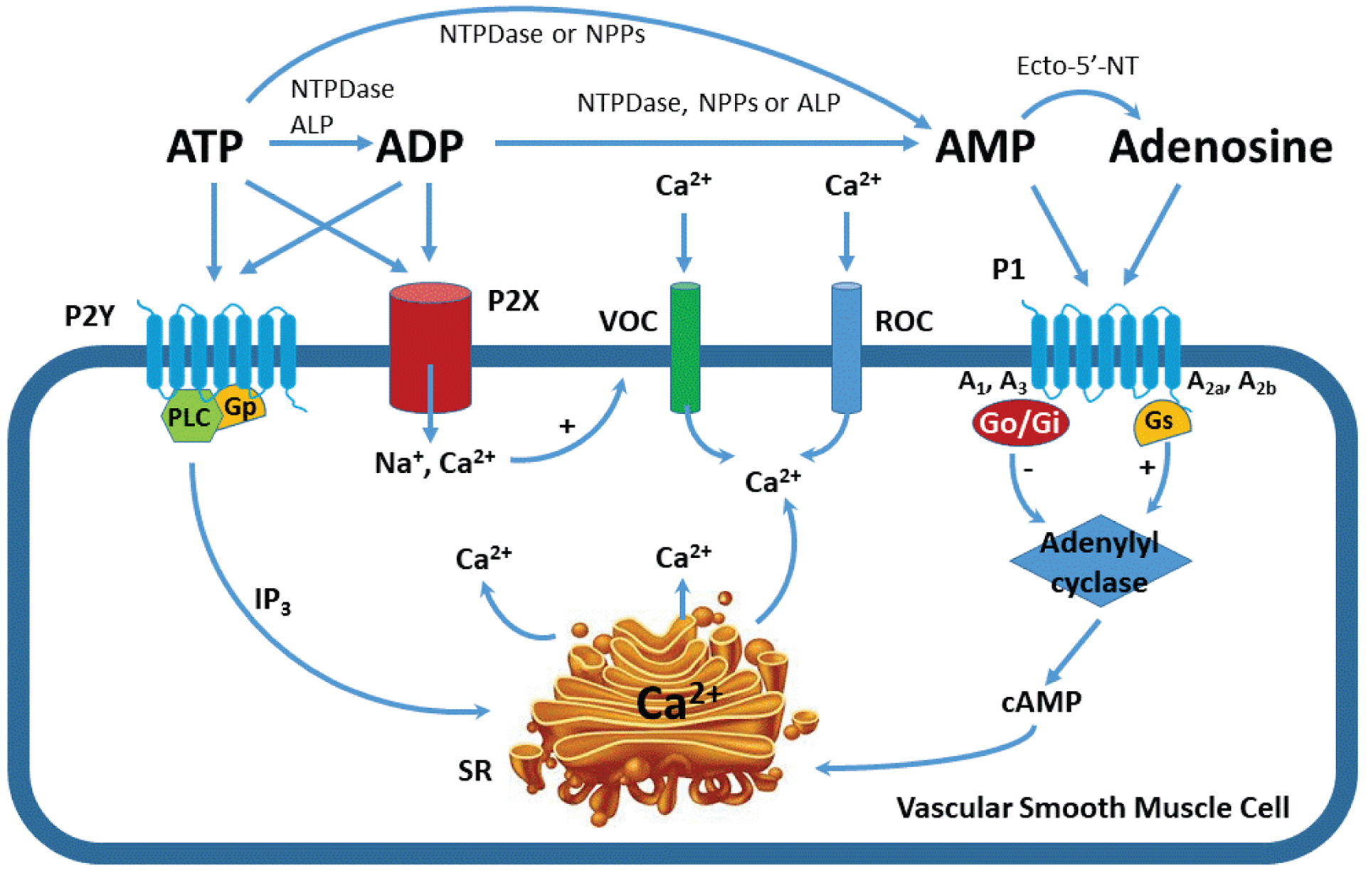
Fig. 1.

Signaling pathways for purinoceptor activation in vascular smooth muscle cells. Illustration of the metabolic pathways for ATP to form ADP, AMP, and adenosine by ectonucleotidases. Adenosine and ATP activate P1 and P2 purinoceptors, respectively. The P1 receptor family is comprised of four subtypes: A1, A2a, A2b and A3. A1 and A3 receptors are linked to a Go/Gi protein and inhibit adenylyl cyclase, thus decreasing cyclic AMP (cAMP). A2a and A2b receptors are linked to Gs protein and stimulate adenylyl cyclase to increase cAMP. The P2 receptor family is divided to two distinct subfamilies: P2X and P2Y. P2X receptors function as non-selective ion-channels whereas P2Y receptors are G protein-coupled receptors. Ecto-5′-NT: ecto-5′-nucleotidases; VOC: voltage-operated calcium channel; ROC: receptor-operated calcium channels; NTPDase: nucleoside triphosphate diphosphohydrolase; NPPs, nucleotide pyrophosphatase/phosphodiesterase; ALP: alkaline phosphatase; IP3: inositol 1, 4, 5-trisphosphate, SR: sarcoplasmic reticulum.
