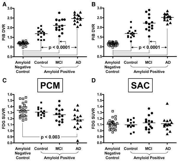
Figure 3.
Group comparisons of PiB retention and glucose metabolism. A–D, PiB retention in bilateral, middle precuneus cortex (A, PCM) or bilateral, subgenual anterior cingulate cortex (B, SAC) and glucose metabolism in PCM (C) or SAC (D) in amyloid-negative controls (open squares), amyloid-positive controls (filled squares), MCI (filled circles), and AD (filled triangles). Significant differences of the amyloid-positive groups compared to the amyloid-negative controls are noted.

