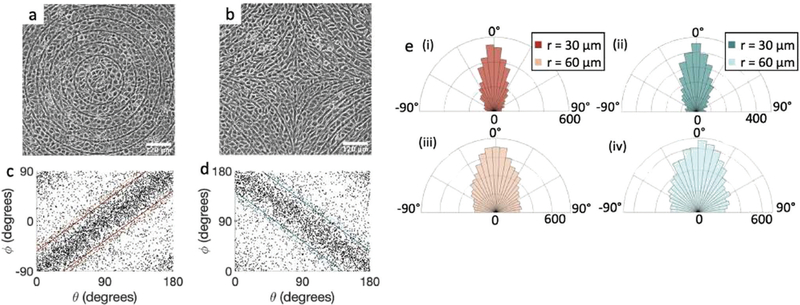
Fig. 3.
Alignment of EpH-4 cells near topological defects. (a) PC of EpH-4 cells in the vicinity of a positive defect on r = 60 μm pattern. Scale bar is 120 μm. (b) PC of EpH-4 cells in the vicinity of a negative defect on r =60 μm pattern. (c) Scatter plot of EpH-4 alignment with r = 30 μm positive defect pattern. Lines of different shades indicate for r =30 μm and r =60 μm. (d) Scatter plot of EpH-4 alignment with r = 30 μm negative defect pattern. Lines of different shades indicate for r = 30 μm and r =60 μm. (e) Alignment of EpH-4 with ridges of (i and ii) r =30 μm and (iii and iv) r =60 μm patterns around (i and iii) positive defects and (ii and iv) negative defects. (i) n = 4 samples and . (ii) has n = 3 and . (iii) has n = 5 and . (iv) has n = 5 and .

