Figure 4.

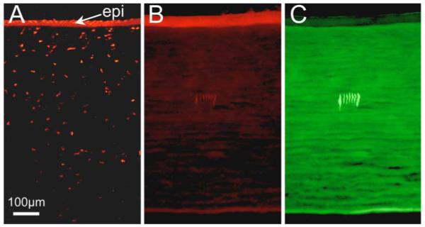
TUNEL assay of cat cornea infused with 1.5% Na-Fl in preservative. (A) Photomicrograph of peripheral portion of a corneal piece showing the segment crushed with a pair of forceps. Note the marked thinning and disruption of the epithelium (epi), along with multiple TUNEL-positive (red fluorescent) cells in both the epithelium and stroma at the crush location. (B) Central portion of the same corneal piece showing TUNEL staining around the seven IRIS lines inscribed ~150 μm below the epithelium. The two most peripheral lines were inscribed at 0.1 mm/s. The middle five lines were inscribed at 2 mm/s. Note the complete absence of TUNEL-positive cells within and around the IRIS lines. (C) Portion of cornea imaged in (B) but shown under 480-nm fluorescent illumination to illustrate that Na-Fl (which fluoresces green under such excitation settings) had fully penetrated the corneal tissue, labeling the stroma uniformly. In contrast, the epithelium shows minimal uptake and fluorescence.
