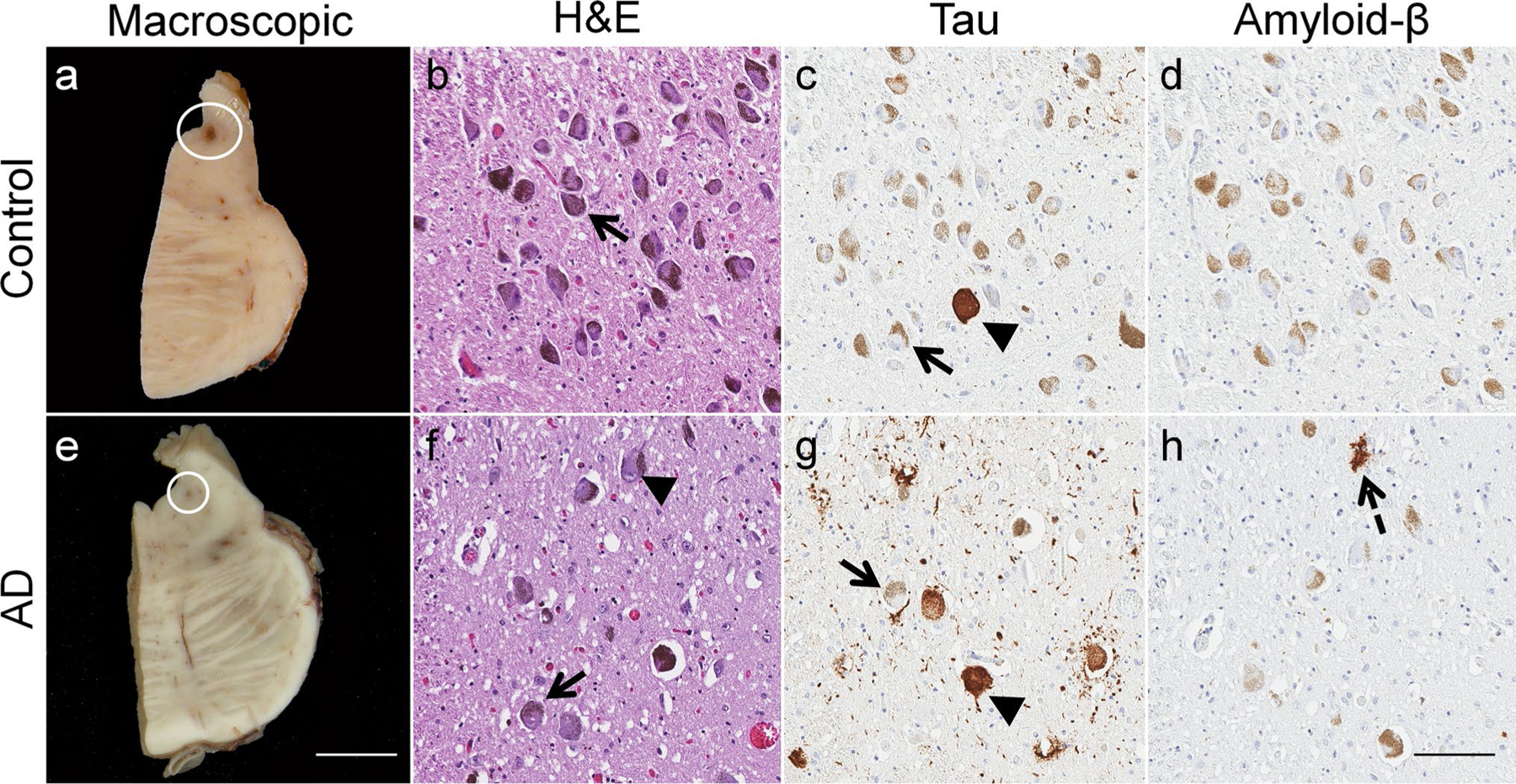
Fig. 3.

Histopathologic changes in the locus coeruleus during Alzheimer’s disease. a Macroscopic examination of the locus coeruleus in an 81-year-old nondemented female revealed a darkly pigmented nucleus due to the presence of neuromelanin (circle). b Microscopic inspection of routine hematoxylin and eosin (H&E) stained section demonstrates a well-populated nucleus with neuromelanin-containing neurons (arrow). c Isolated neurofibrillary tangles (arrowhead) were identified using tau immunohistochemistry (dark brown), which contrasts with the more yellow–brown appearance of healthy, neuromelanin-containing neurons (arrow). d No amyloid deposits were identified in the nondemented control. e Macroscopic examination of the brain of an 80-year-old female neuropathologically diagnosed with Alzheimer’s Disease revealed a significantly depigmented locus coeruleus (circle), f reflective of the decrease in the population of pigmented neurons (arrow) observed microscopically on an H&E-stained section. Neurofibrillary tangles visibly displace neuromelanin (arrowhead). g Tau immunostaining reveals neurofibrillary tangles (arrowhead), which contrast in comparison to the yellowish-brown neuromelanin pigment observed in surviving neurons. h An isolated amyloid-β deposit was visible (dotted arrow). Case characteristics—Nondemented control: Braak stage = 0, Thal phase = 0; Sporadic Alzheimer’s disease: Braak stage = V, Thal phase = 5; Immunohistochemistry—Tau marker: PHF-1, Amyloid-β marker: 6F/3D; Scale bar— Macroscopic = 8 mm; Microscopic—100 μm
