
Figure 3.
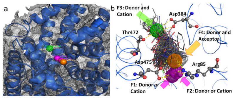
Pharmacophore query for the vestibular DAT binding pocket obtained by inhibitor and substrate docking. Panel a: Five pharmacophoric features were created: F1 and F2, donor/cation (pink spheres); F3, donor/cation (green wire mesh sphere); F4, donor/acceptor (orange sphere); F5, excluded volume (grey wire mesh spheres). Panel b: The amino acids with potentiality for ligand binding (Arg85, Asp384, Thr472 and Asp475) and their spatial relationship with the pharmacophoric features are depicted as atom colored sticks. Possible H-bond interactions with pharmacophore features are depicted as yellow dashed lines.