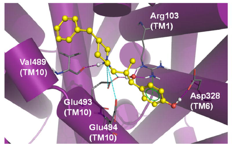
Figure 9.

MI-4 docking in the SERT model. Optimal positioning of MI-4 in the SERT vestibular pocket is depicted by the top-ranked pose generated by MOE-Dock 2007. Ligand pocket side chains (atom colored) within 5 Å of the docked MI-4 molecule (yellow / atom colored) are shown. Intermolecular H-bond (pink dashed lines), π-cation (green dashed lines) and ionic (cyan dashed lines) interactions are indicated.
