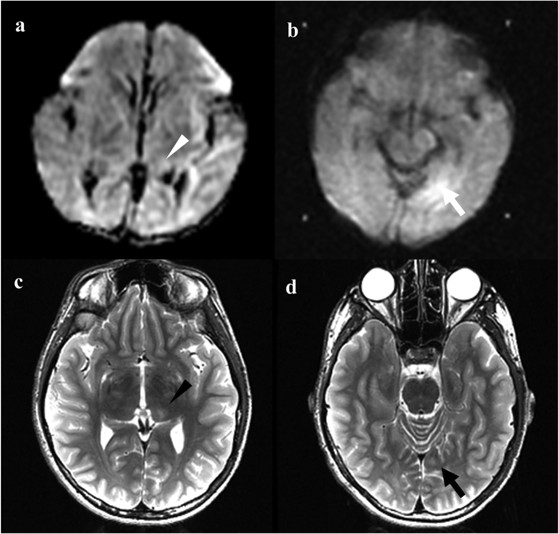
Figure 3.

Neonatal and adolescent magnetic resonance imaging (MRI) of patient no 8. a Axial diffusion weighted imaging of the neonatal brain demonstrates reduced diffusion in the left thalamus (white arrowhead). b Additional area of reduced diffusion in a watershed distribution in the medial left occipital lobe on neonatal MRI (white arrow). c Axial T2-weighted imaging in adolescent MRI demonstrates T2 hyperintensity and volume loss in the left thalamus (black arrowhead), corresponding to area of reduced diffusion in a. d T2 hyperintensity in the medial left occipital subcortical white matter (black arrow) corresponding to area of reduced diffusion in b.
