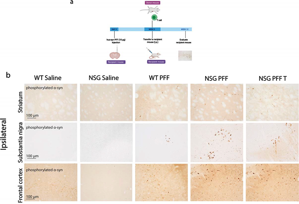
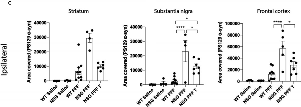
Figure 2. Reduced phosphorylated α-syn pathology in immunocompromised mice that received adoptive transfer of T cells.
a. Timeline of experiment. b. Phosphorylated α-syn was detected in the ipsilateral hemisphere to PFF injection in the striatum, substantia nigra and frontal cortex. The reconstitution of T cells was conducted in two separate experiments and the results pooled after results of the reduction in phosphorylated α-syn were shown to be consistent between the two experiments. c. Densitometry of 5–9 mice per group to determine the area covered in phosphorylated α-syn levels in the ipsilateral striatum, substantia nigra and frontal cortex. Wildtype Saline, n=5; NSG Saline n=5, wildtype PFF, n=9; NSG PFF, n=5; NSG PFF T n=7). d. Statistical analyses were performed by Kruskal-Wallis test * p<0.05, **** p<0.001. Scale bar: 100 μm. Schematic created with BioRender.com


