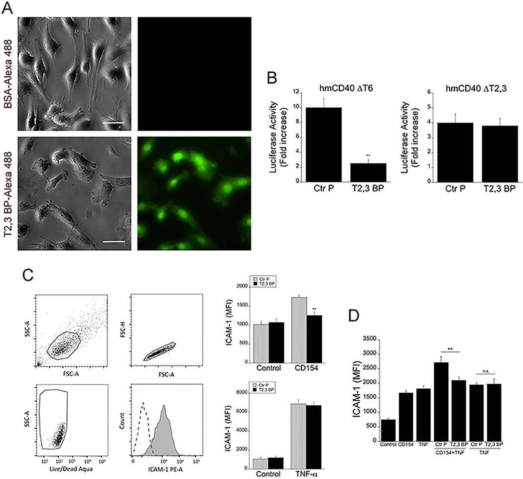
Figure 1. ri CD40-TRAF2,3 blocking peptide penetrates cells, inhibits CD40-TRAF2,3 signaling and impairs CD40-driven ICAM-1 upregulation.
A, Human Müller cells were incubated in medium containing Alexa Fluor 488-conjugated ri CD40-TRAF2,3 blocking peptide (T2,3 BP) or Alexa Fluor 488-conjugated bovine serum albumin (BSA; both at 10 μM) for 3 h. Scale bar, 50 μm. Original magnification X400. Images represent fluorescence of unfixed Müller cells after extensive washing of monolayers. B, Mouse endothelial cells (mHEVc) that express an NF-κB response element that drives transcription of a luciferase reporter plus either hmCD40 ΔT2,3 or hmCD40 ΔT6 were pre-incubated with ri control peptide (Ctr P) or ri CD40-TRAF2,3 blocking peptide (T2,3 BP; both at 1 μM) or medium alone followed by stimulation with human CD154. Data are expressed as fold-increase in normalized luciferase activity in cells stimulated with CD154 compared to cells treated with respective peptide in the absence of CD154. C, Human retinal endothelial cells were treated with ri control peptide (Ctr P) or ri CD40-TRAF2,3 blocking peptide (T2,3 BP; both at 1 μM) followed by stimulation with CD154 or TNF-α (100 pg/ml) for 24 h. Expression of ICAM-1 was assessed by flow cytometry. Dot plot and histogram show gating strategy. ICAM-1 was analyzed on live cells that did not stain with Aqua LIVE/DEAD kit. D. Human retinal endothelial cells were incubated with or without TNF-α (30 pg/ml) followed by treatment with peptides and stimulation with CD154. Data shown represent mean ± SD of triplicate samples. Results are representative of 3 independent experiments. **P<0.01 by ANOVA.

