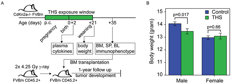
Figure 1. THS exposure altered body weight of three-week-old male pups compared to control.
A. Study design. Mice were exposed to THS starting from the first day of pregnancy (post coital, p.c.) until the pups were weaned at 3-weeks of age. Plasma cytokine levels were measured at 2 days of age. Body weight was assessed at weaning. Bone marrow (BM), spleen (SP) and peripheral blood (PB) were collected at 5 weeks of age for immunophenotyping and bone marrow was transplanted into irradiated recipients. Tumor development was monitored for one year.
B. Bars represent body weight (gram) at weaning for control and THS exposed male and female mice [n=142 pups (19 litters) in the Control group and 105 pups (15 litters) in THS-treated group]. Data are presented as the mean and error bars indicate standard error. P-values were obtained using the two-tailed t-test.

