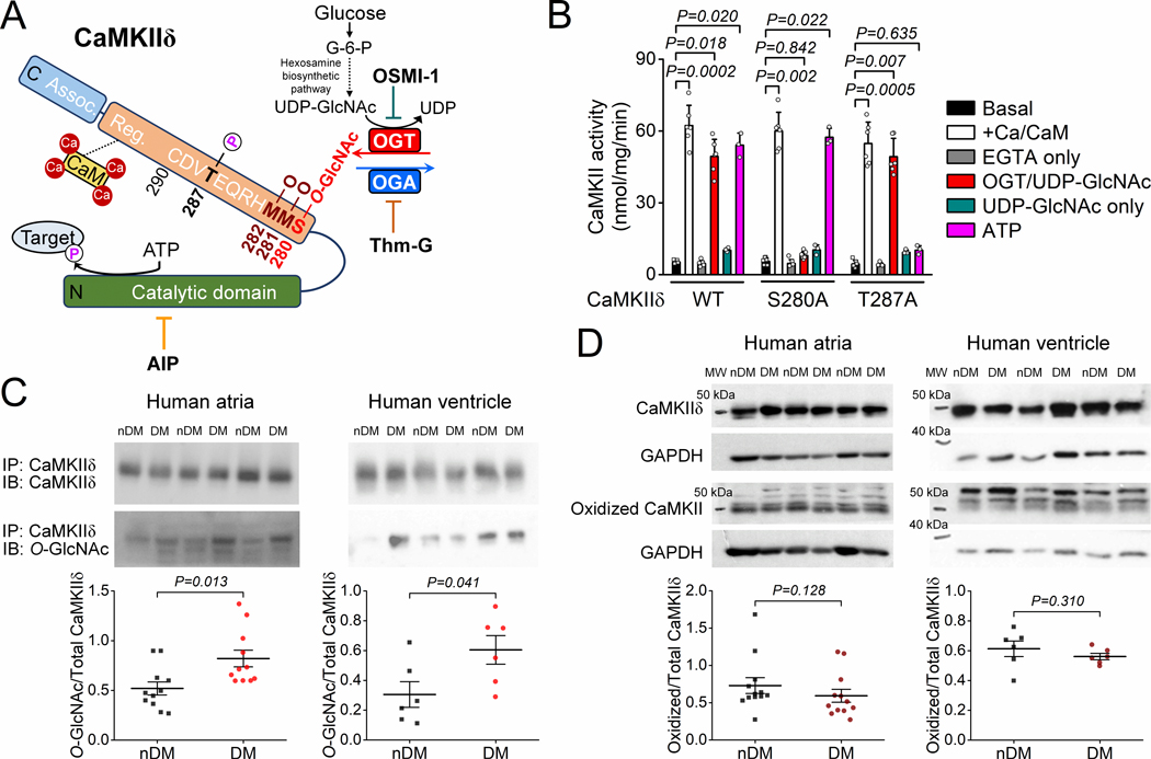
Figure 1. CaMKIIδ O-GlcNAcylation is increased in human diabetic hearts.
A, Schematic of CaMKIIδ structure and sequence of the regulatory hub domain and the O-GlcNAcylation pathway. B, CaMKII activity measured as 32P incorporation in HEK293 cell lysates expressing WT, S280A, and T287A CaMKIIδ (n, basal=6, Ca/CaM=6, EGTA=6, OGT/UDP-GlcNAc=6, UDP-GlcNAc=3, ATP=3 in all three CaMKIIδ forms). Kruskal-Wallis one-way ANOVA, followed by Dunn’s multiple comparisons test. C, Western blot data showing enhanced CaMKII O-GlcNAcylation in human atrial and ventricular samples from diabetic (DM) versus non-diabetic (nDM) patients (n, atrial-DM=11, atrial-nDM=11, ventricular-DM=6, ventricular-nDM=6). Mann-Whitney test. D, CaMKII oxidation was not increased in atrial and ventricular samples from patients with diabetes (n, atrial-DM=12, atrial-nDM=12, ventricular-DM=6, ventricular-nDM=6). Mann-Whitney test. MW indicates molecular weight.

