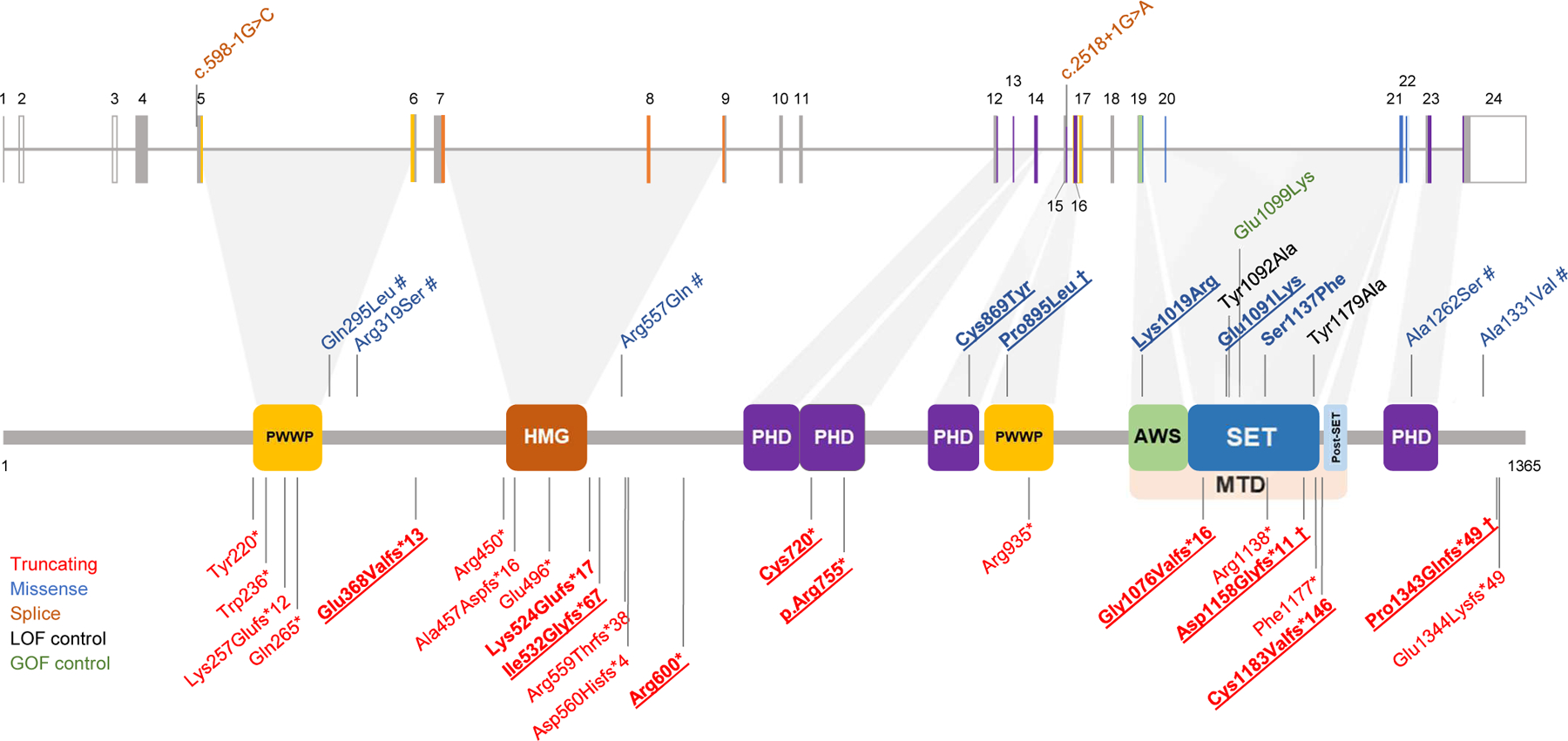
Figure 1. Localization of the NSD2 variants.

The diagram shows the structure of the NSD2 gene (above) and protein (below, isoform 1, encoded by the NM_133330.2 transcript) together with the variants discussed in this study as well as variants that have previously been reported in large sequencing studies. The variants carried by the 18 additional individuals described in this study are in bold. Observed germline variants absent in the HGMD database are underlined. † = shared by more than one patient; # = VUS/likely benign. The observed pathogenic missense variants we found map to three distinct domains of NSD2: a PHD zinc-finger domain (residues 831–875), a PWWP domain (residues 880–942) and the catalytic methyltransferase domain (residues 1011–1203; composed of three subdomains, namely an AWS, a SET and a post-SET domain). The colour coding of the variants and protein domains is reported in the legend. PWWP: proline–tryptophan–tryptophan–proline domain (IPR000313); HMG: high mobility group box domain (IPR009071); PHD: zinc-finger domain, Plant-HomeoDomain type (IPR001965); AWS: Associated With SET domain (IPR006560); SET: Su(Var)3–9, enhancer-of-zeste, trithorax domain (IPR001214); MTD: catalytic methyltransferase domain, composed by the AWS, SET and a post-SET domain. Domains were annotated according to the Uniprot databank (see Web Resources).
