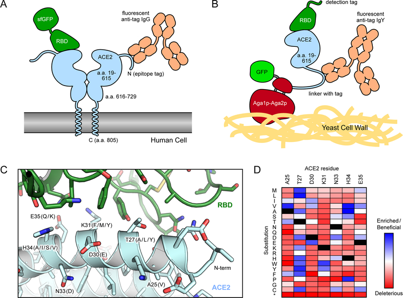
Figure 2. Selection strategies for enhancing the affinity of ACE2 for the RBD of SARS-CoV-2.
(A) In human cell selections of ACE2 variants, surface expression of full-length ACE2 with N-terminal epitope tags was detected via fluorescent anti-tag antibodies. Cells expressing ACE2 clones with high binding to RBD-sfGFP were collected by FACS. (B) In FACS selections of yeast, the protease domain of ACE2 was expressed as a fusion to Aga2p for display on the yeast surface. Displayed protein was detected via a C-terminal fusion to GFP or immunostaining of an epitope tag in the connecting linker. Bound RBD was labeled via biotin or an epitope tag for fluorescence detection. (C) Structure (PDB 6M17) of RBD (green) bound to ACE2 (pale blue). Common mutations in high affinity engineered decoys are indicated in parentheses. (D) Mutational landscape showing how single amino acid substitutions in ACE2 increase (blue) or decrease (red) binding to RBD. Data are from Chan et al50.

