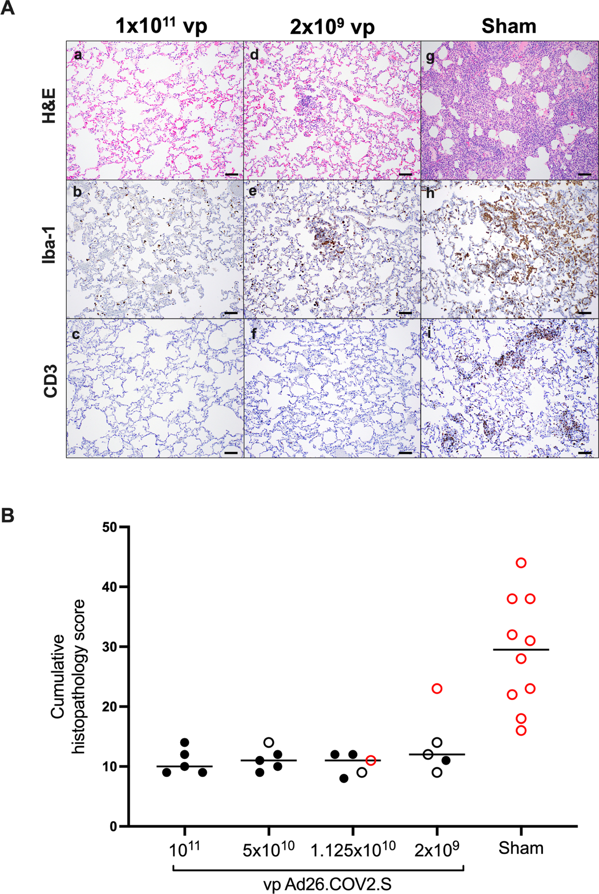
Figure 7. Comparative pathology in vaccinated and unvaccinated animals following SARS-CoV-2 challenge.

(A) Representative pathology from animals vaccinated with (a–c) 1×10¹¹ vp Ad26.COV2.S, (d–f) 2×10⁹ vp Ad26.COV2.S, or (g–i) sham negative controls on day 10 following SARS-CoV-2 challenge. (a, d, g) representative H&E histopathology. (b, e, h) immunohistochemistry for Iba-1 (macrophages). (c, f, i) showing immunohistochemistry for CD3 (T-lymphocytes). Animals that received a high vaccine dose had minimal evidence of SARS CoV-2 pathology (a–c). Animals that received the lowest vaccine dose showed focal pathology (d–f) characterized by increased alveolar macrophages, focal interstitial septal thickening and aggregates of macrophages. Sham vaccinated animals had locally extensive moderate interstitial pneumonia (g) characterized by extensive macrophage infiltrates (h) and expansion of perivascular and interstitial CD3 T lymphocytes (i). (B) Histopathologic scoring of lung lesions in all lobes in vaccinated and sham animals following SARS-CoV-2 challenge. Scoring was performed independently by two blinded, veterinary pathologists. Lesions reported included 1) inflammation interstitial/septal thickening 2) infiltrate, macrophage 3) alveolar infiltrate, mononuclear 4) perivascular infiltrate, macrophage 5) bronchiolar type II pneumocyte hyperplasia 6) BALT hyperplasia 7) inflammation, bronchiolar/peribronchiolar infiltrate 8) neutrophils, bronchiolar/alveolar and 9) infiltrate, eosinophils. Lesions such as focal fibrosis and syncytia were reported but not included in scoring. Edema, alveolar flooding was excluded from scoring since animals received terminal bronchoalveolar lavages. Each feature assessed was assigned a score of 0= no significant findings; 1=minimal; 2= mild; 3=moderate; 4=marked/severe. Eight representative samples from cranial, middle, and caudal lung lobes from the left and right lungs were evaluated from each animal and were scored independently. Scores were added for all lesions across all lung lobes for each animal for a maximum possible score of 288 for each monkey. Lungs evaluated were inflated/suffused with 10% formalin. Horizontal lines reflect median values. Solid black circles indicate animals that showed no virus in BAL and NS following challenge, open black circles indicate animals that showed virus in NS but not BAL following challenge, and open red circles indicate animals that show virus in both BAL and NS following challenge. Scale bars = 100 microns.
See also Figures S5, S6 and Table S1.
