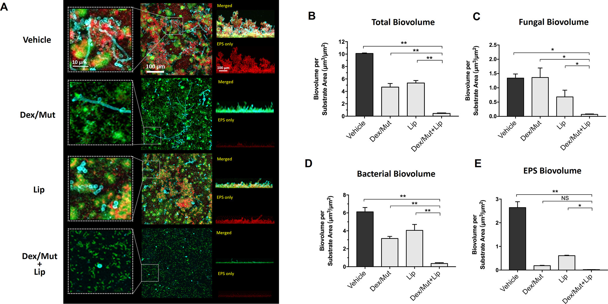
Figure 4: Prevention of fungal-bacterial mixed biofilm by topical sequential treatment of commercial Lipase and Dextranse/Mutanse combination.

Commercial enzymes of the optimum activity units for (1000 U/mL for lipase and 525/105 U/mL for dextranase/mutanase, respectively) were used. (A) Three-dimensional confocal images of the fungal-bacterial mixed biofilm formed after the topical sequential treatments. C. albicans cells (yeasts or hyphae) are depicted in cyan; S. mutans cells are depicted in green; The EPS glucan matrix is depicted in red. Representative merged biofilm images are displayed in the middle panel, while a magnified (close-up) view of each small box is positioned in the left panel. Lateral (side) views of each biofilm are displayed at the right panel (the merged image at the top and the EPS channel at the bottom). (B-E) Quantitative computational analysis of the confocal images. The title of each graph indicates the channel(s) used for individual analysis. *, p<0.05; **, p<0.01 (1-way analysis of variance with Tukey’s multiple comparisons test). Enzyme unit of lipase and dextranase/mutanase represent μmol of pNP and reducing sugar produced in 1 hour, respectively.
