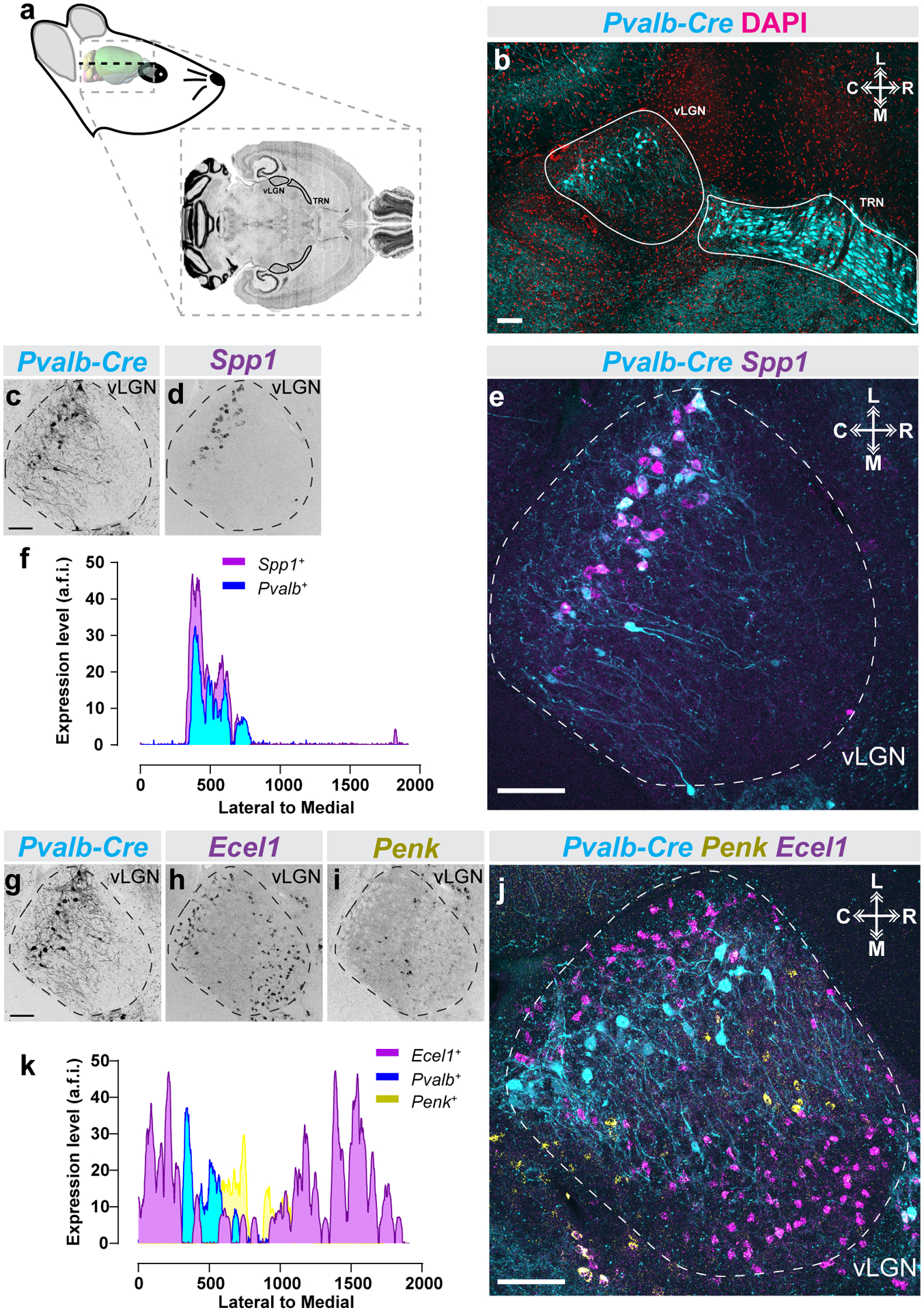
Figure 5. Subtype specific laminar organization of vLGN neurons along the rostro-caudal axis.

(a) Schematic of horizontal mouse brain sectioning used to find vLGN. (b) Identification of the mouse vLGN and thalamic reticular nucleus (TRN) in the horizontal plane using the Pvalb-Cre::Thy1-Stop-YFP transgenic reporter and DAPI counter-staining. (c-e) ISH-labeling for Spp1+ neurons in horizontal vLGN of P60 Pvalb-Cre::Thy1-Stop-YFP transgenic reporter mice. (f) Line scan analysis of spatial distribution of Spp1+ and Pvalb+ cells. Arbitrary fluorescence units (a.f.i.) are plotted against distance from the lateral-most part of the vLGN to the most medial. (g-j) Double ISH for Ecel1 and Penk mRNA in horizontal vLGN of P60 Pvalb-Cre::Thy1-Stop-YFP transgenic reporter mice. (k) Line scan analysis of spatial distribution of Ecel1+, Penk+, and Pvalb+ cells in vLGN, plotted as in (f), revealing four discrete spatial domains of vLGN. n=1 animal for all data presented. All scale bars = 100 μm.
