Figure 3:
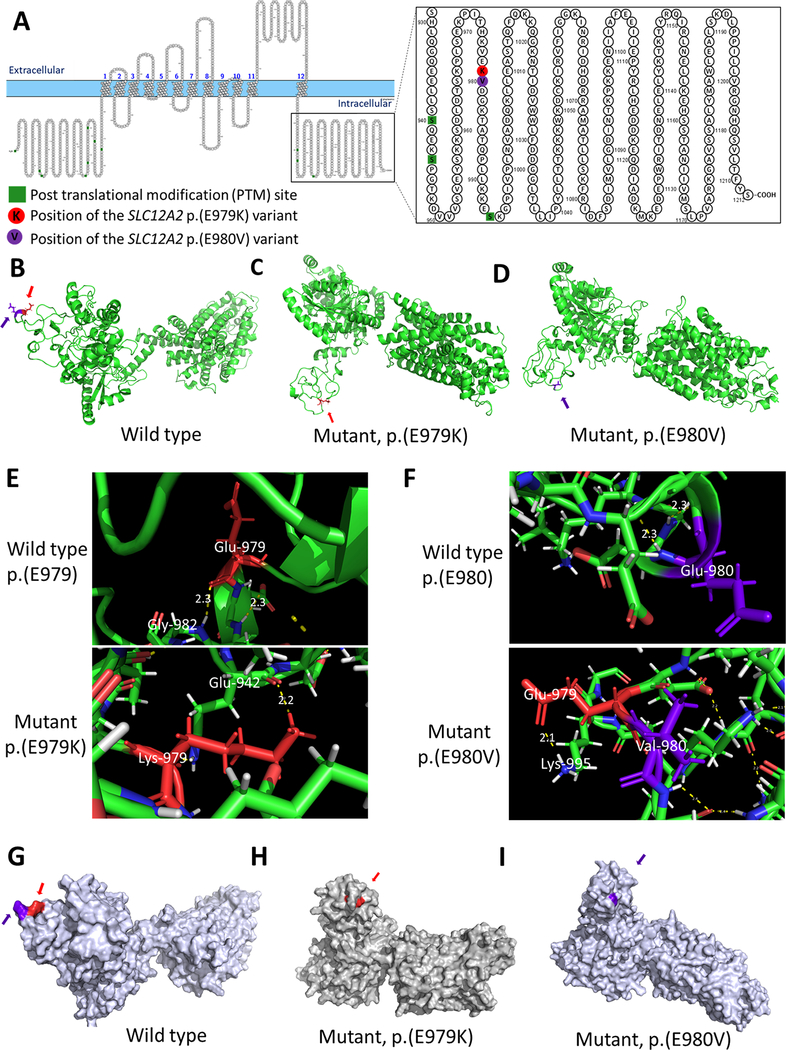
SLC12A2 protein modelling. (A) Primary structure of SLC12A2 protein showing the site of the p.(E979K) and p.(E980V) variants. The ribbon protein model of the (B) wild type, (C) mutant p.(E979K), and (D) mutant p.(E980V) proteins. (E) The variant position showing wild-type residue (E979) and mutant (K979) hydrogen bonds. (F) The variant position showing wild-type residue (E980) and mutant (V980) hydrogen bonds. The surface protein model of the (G) wild type, (H) mutant p.(E979K), and (I) mutant p.(E980V) proteins. The site of the p.(E979K) variant is highlighted in red (pointed to by the red arrows) and the site of the p.(E980V) variant is highlighted in violet (pointed to by the violet arrows).

