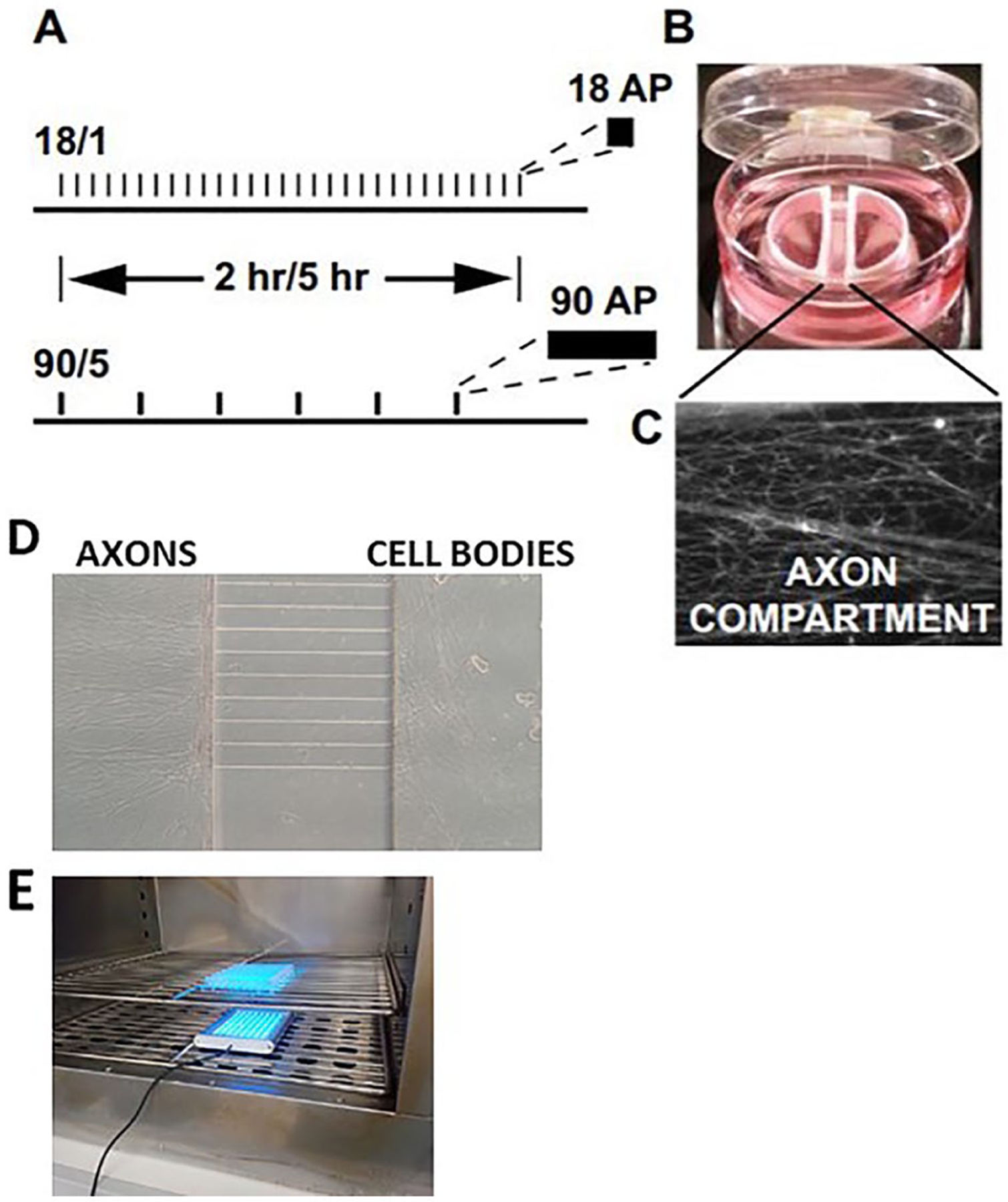
Figure 1.

Cell culture preparations used for the study of gene expression in dorsal root ganglion (DRG) neurons in response to action potential firing patterns. Action potentials delivered at a frequency of 10 Hz in 1.8-second bursts, repeated at 1-minute intervals, or stimulated in 9-second bursts, repeated at 5-minute intervals (termed 18/1 and 90/5, respectively) (A). Stimulation was delivered for 2 or 5 hours for both 18/1 and 90/5 stimulation patterns, resulting in an equal number of electrical pulses delivered by bipolar stimulating electrodes across a Campenot chamber. In (B and C), a representative image of a Campenot chamber used for culture of DRG neurons showing axonal outgrowth (C) in the central compartment. Platinum electrodes deliver biphasic stimulation to both cell body compartments. Reprinted from Lee and others (2017). (D) Custom-made microfluidic chamber used for electrical or optogenetic stimulation showing DRG cell bodies and axons. (E) Optogenetic stimulation of DRG neurons in cell culture incubator.
