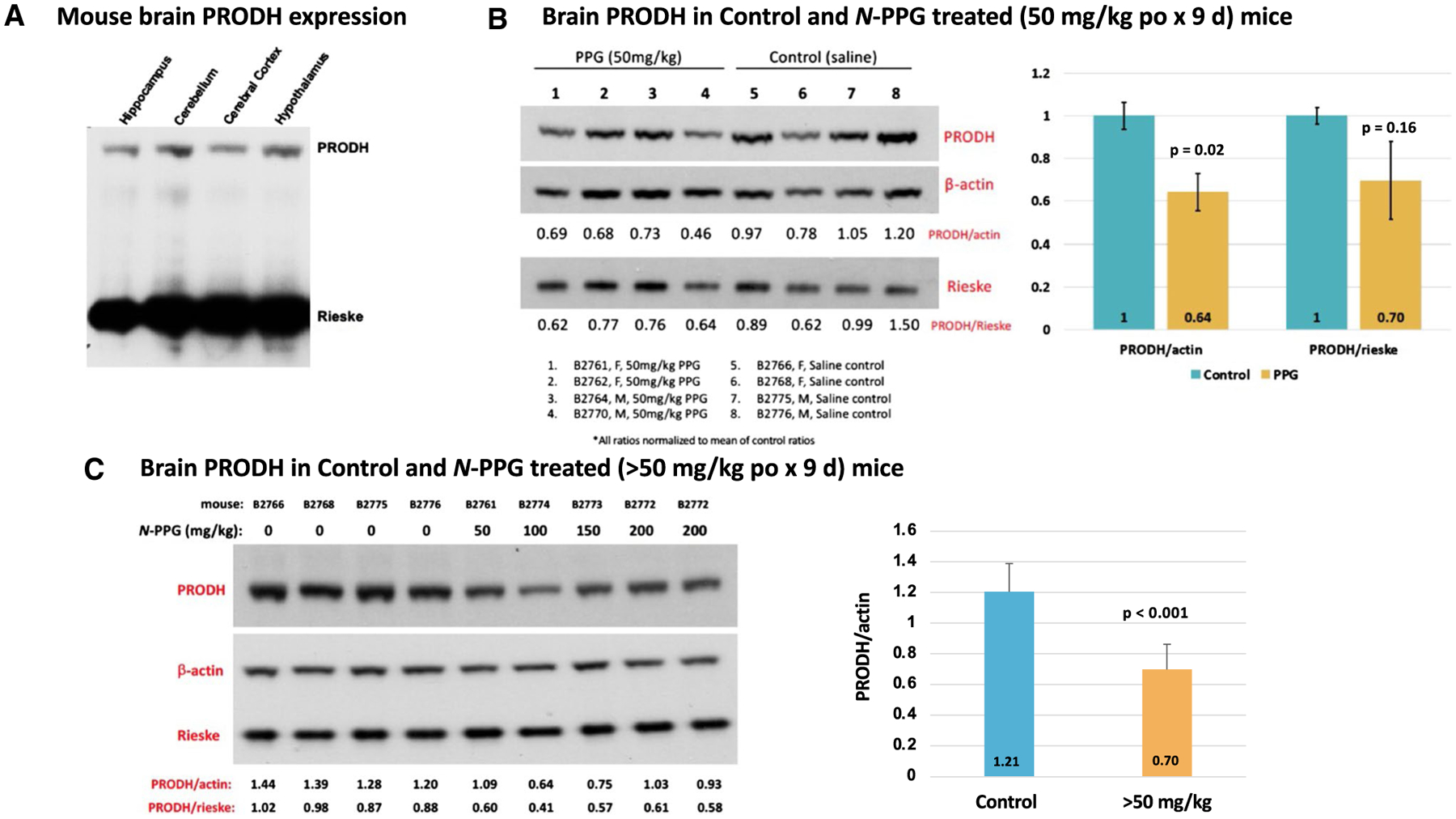
Fig. 3.

PRODH expression in mouse whole brain samples and its reduction following 9 days of oral N-PPG treatment. A Commercially obtained lysates of indicated mouse brain tissue samples immunoblotted for PRODH protein expression, with total gel protein load normalized to mitochondrial Rieske content. B Western blots of whole brain samples from wildtype B6/CBA mice (5 days old) orally treated for 9 days with either vehicle (saline) or 50 mg/kg N-PPG (left panel); image analysis quantification of probed PRODH signals normalized to either β-actin or Rieske, with bar graphs of mean ratio values (± SD) showing at least 30% reduction in mean PRODH expression following N-PPG treatment (right panel, ANOVA F test p values). C. Western blots loaded with whole brain lysates from four mice orally treated with saline (0 mg/kg N-PPG), one with 50 mg/kg N-PPG, and three with > 50 mg/kg N-PPG treatments (100, 150, 200 mg/kg), with an extra lane loaded with lysate from the highest dose-treated sample (left panel). Independent technical replicates of this 9-lane western blot format each probed for PRODH, β-actin and Rieske were scanned and their calculated PRODH ratios compared (right panel) to demonstrate a > 40% reduction in PRODH expression across the > 50 mg/kg treated samples relative to all saline-treated control samples (ANOVA F test p value). Western blot molecular weight standards confirmed protein running sizes as indicated in Fig. 2: PRODH at 68 kDa, β-actin at 42 kDa, and Rieske at 30 kDa
