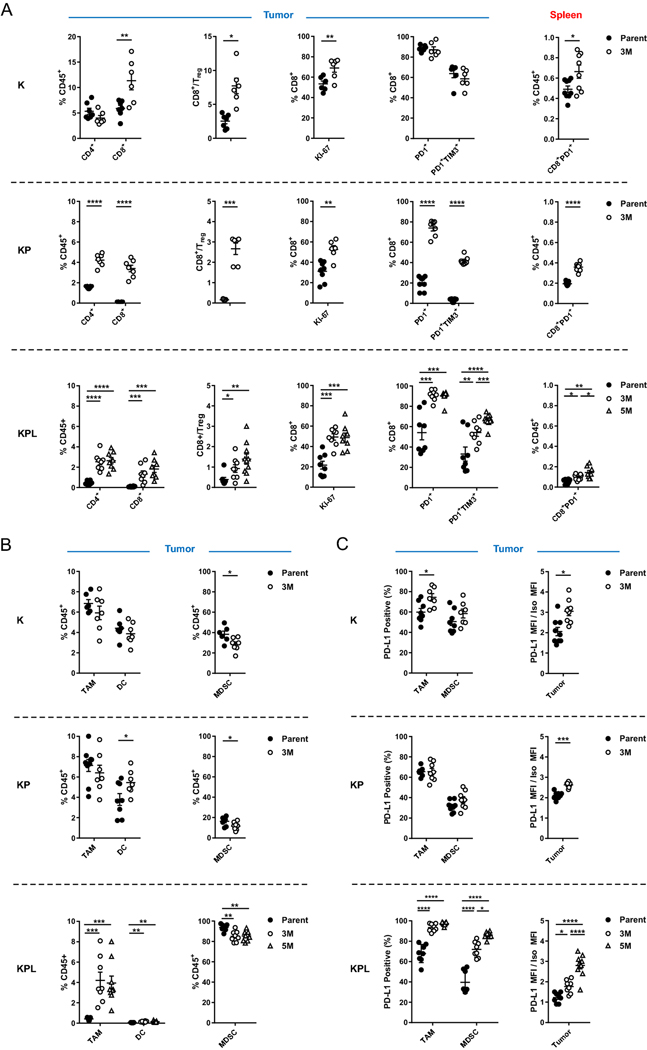
Fig. 3.
Distinct immune phenotypes of murine models revealed by FACS. On day 14–16 post-tumor inoculation [2 × 10⁶ K-Parent and K-3M cells in 129-E mice; 8 × 10⁵ KP-Parent and 2 × 10⁶ KP-3M cells in FVB mice; 1 × 10⁵ KPL-Parent, 1.5 × 10⁵ KPL-3M, and 3 × 10⁵ KPL-5M cells in FVB mice], tumors and spleens were harvested and analyzed by FACS. a Lymphoid compartment. b Myeloid compartment. c PD-L1 expression. Data are representatives of at least two biological replicates of 6–10 mice per group. P values were determined by P values were determined by two-tailed non-paired Student’s t test. *P < 0.05; **P < 0.01; ***P < 0.001; ****P < 0.0001

