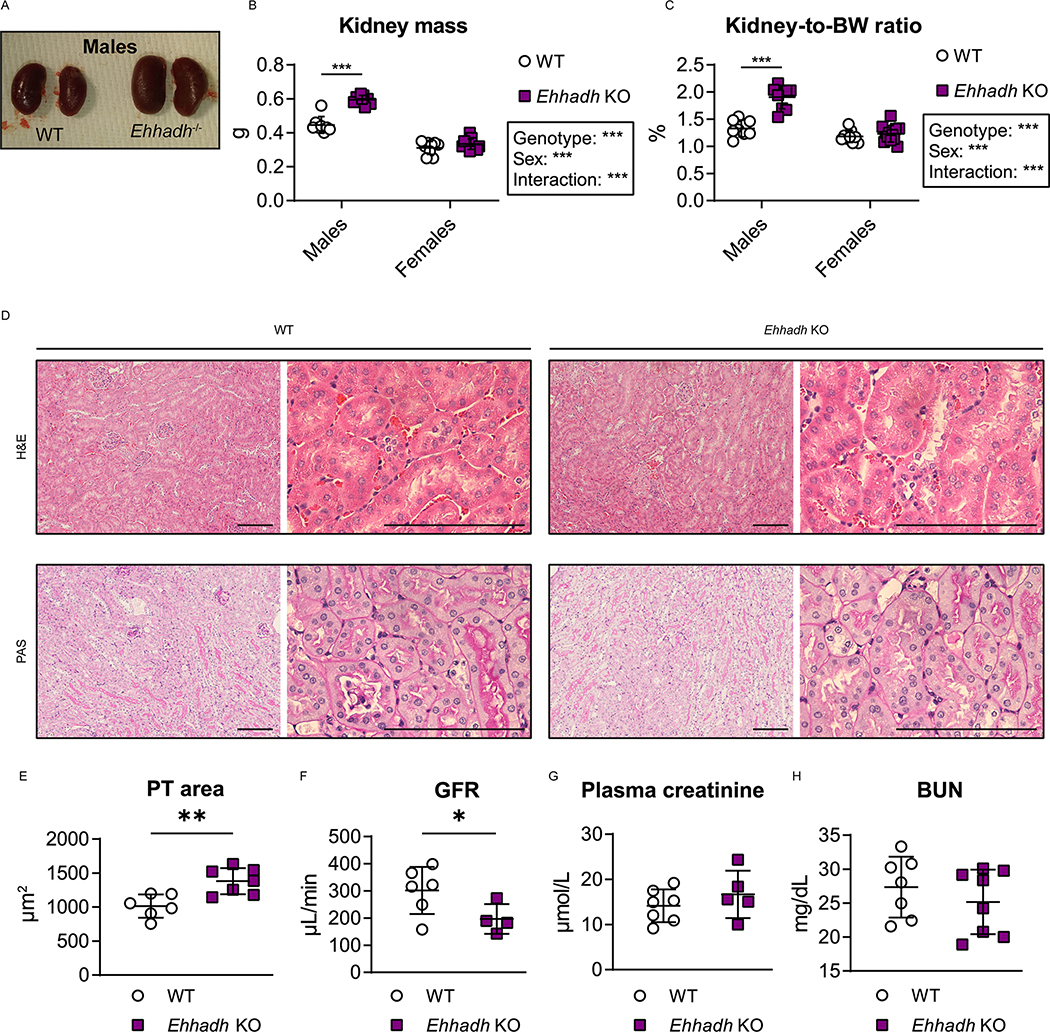
Figure 1. EHHADH deficiency induces male-specific kidney hypertrophy without signs of severe pathology.
A) Representative images of WT and Ehhadh KO kidneys from male mice. B, C) Measurement of B) kidney weight (combined weight of both kidneys per animal) and C) kidney-to-body weight (BW) ratio in 4 to 9 month-old male WT (n=8), male Ehhadh KO (n=9), female WT (n=9), and female Ehhadh KO mice (n=12). D) Representative images of H&E and PAS staining of kidney sections from male WT and Ehhadh KO mice. Scale bars = 100 μm. E) Morphometric analysis of the cross-sectional tubule areas in WT (n=6) and Ehhadh KO (n=7) male mice F) Glomerular filtration rate (GFR) in WT (n=6) and Ehhadh KO (n=4) male mice. G) Plasma creatinine levels (μmol/L) in WT (n=5) and Ehhadh KO (n=7) male mice. H) Blood urea nitrogen (BUN) levels (mg/dL) in WT (n=7) and Ehhadh KO (n=8) mice male. Data are presented as mean ± SD with individual values plotted. ***P < 0.001, **P < 0.01, *P < 0.05, by two-way ANOVA (B and C), or unpaired t-test with Welch’s correction (E, F).

