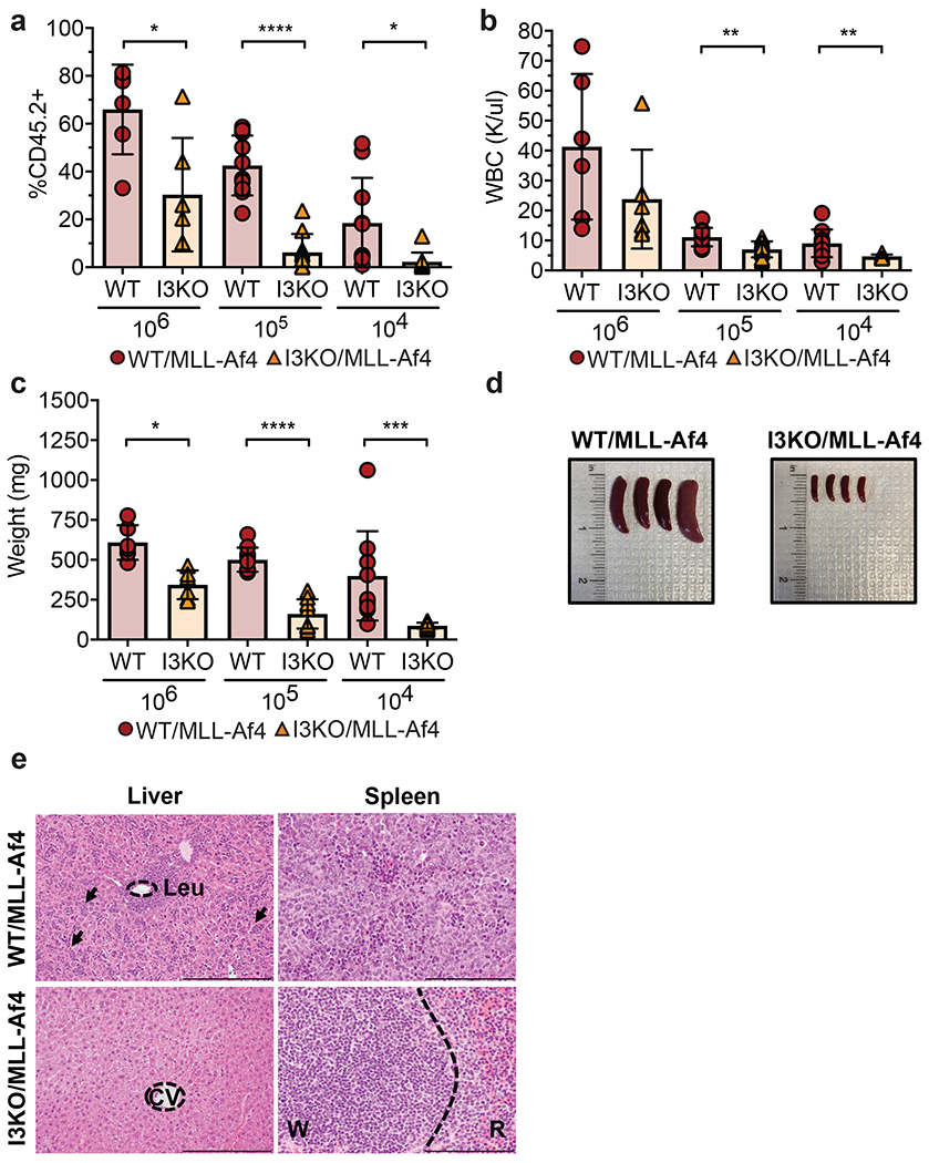
Figure 4: Igf2bp3 deletion is necessary for MLL-Af4 leukemia-initiating cells to reconstitute mice in vivo.

a) Percentage of CD45.2+ in the peripheral blood of secondary transplanted mice from leukemic WT/MLL-Af4 or I3KO/MLL-Af4 donor mice at 106, 105, and 104 BM cells at 4 weeks post-transplantation (For all panels in this figure: n=6 recipient mice per genotype for 106 cells and n=10 recipient mice per genotype for 105 and 104 cells; t-test; *P < 0.05, ***P < 0.001, ****P < 0.0001).
b) WBC from PB of secondary transplanted mice from WT/MLL-Af4 or I3KO/MLL-Af4 BM 3–4 weeks post-transplant (t-test; **P < 0.01).
c) Splenic weights of secondary transplanted mice at 4-5 weeks (t-test; *P < 0.05, ***P < 0.001, ****P < 0.0001).
d) Images of splenic tumors in secondary mice transplanted with 10,000 BM cells from WT/MLL-Af4 mice (left) or I3KO/MLL-Af4 mice (right) at 5 weeks.
e) H&E staining of liver and spleen of secondary transplant recipients that received 105 cells at 4 weeks. Scale bar: liver, 200 microns; spleen, 100 microns; CV=Central vein; W=White pulp; R=Red pulp; Leu= Leukemia; arrows showing infiltration.
