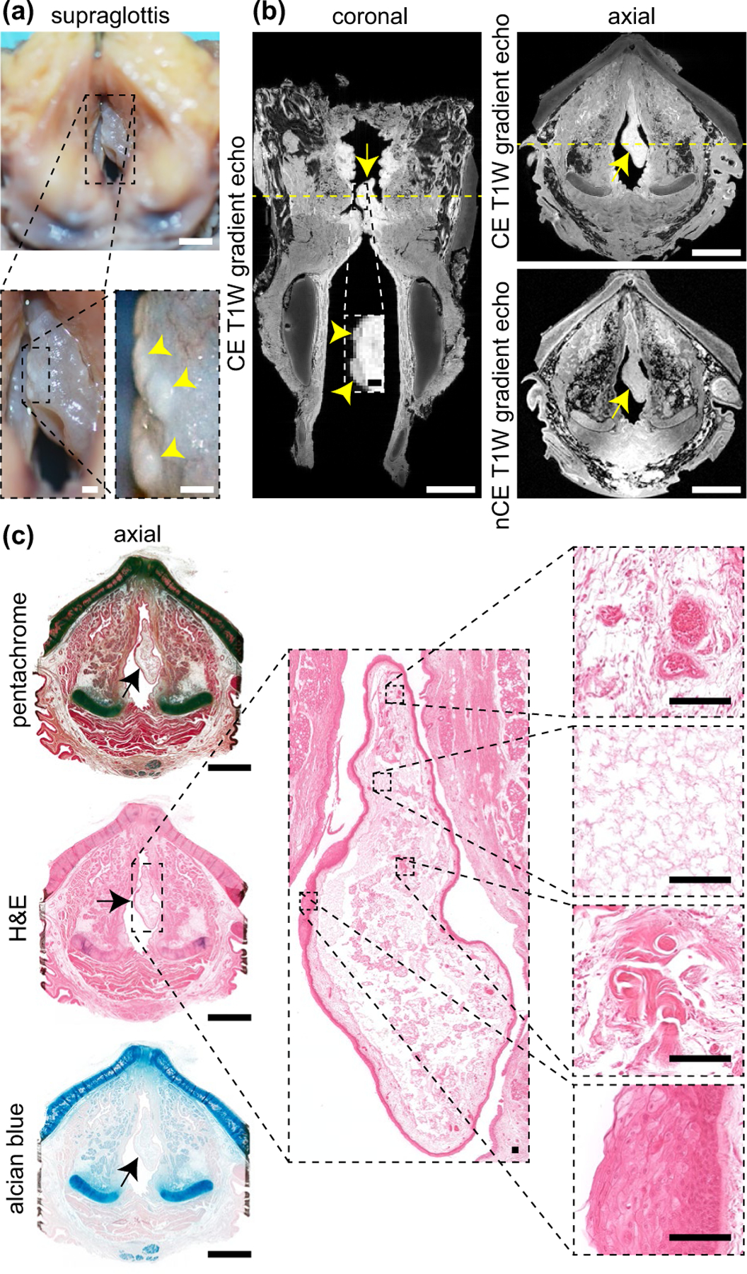
FIGURE 3.

MRI and histological characterisation of polypoid degeneration. (a) Superior view of the supraglottis and vocal fold mucosae at the time of procurement from a 74-year-old female donor. Yellow arrowheads denote leukoplakia on the epithelial surface. (b) CE (coronal and axial) and nCE (axial) T1W gradient echo images showing the lesion boundary in both planes. Yellow arrows denote the lesion; yellow arrowheads denote leukoplakia. Dashed yellow lines denote the position of orthogonal views in the respective CE coronal and axial images. Note: The lateral thyroid cartilage laminae were resected prior to imaging. (c) Pentachome-, H&E- and alcian blue-stained histological images of the lesion in the same axial view as shown on MRI. Black arrows denote the lesion. The high-magnification H&E images show (top-to-bottom): capillary wall thickening and extravascular leakage of red blood cells; an edematous lake; dense fibrin deposits; epithelial hyperparakeratosis and basement membrane thickening. CE, contrast-enhanced; H&E, haematoxylin and eosin; nCE, non-contrast-enhanced; MRI, magnetic resonance imaging; T1W, T1-weighted. Scale bars, 5 mm (whole-larynx photography, MRI and histology); 500 μm (high-magnification photography and MRI); 100 μm (high-magnification histology)
