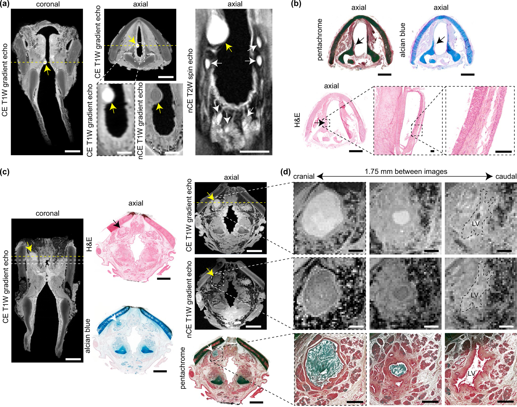
FIGURE 5.

MRI and histological characterisation of mucus retention cysts. (a) CE T1W gradient echo coronal and axial images, alongside high-magnification CE T1W gradient echo, nCE T1W gradient echo, and nCE T2W spin echo axial images, showing an infraglottic lesion in a 10-year-old female. Yellow arrows denote the lesion; white arrows denote adjacent mucous glands within the posterior infraglottis. Dashed yellow lines denote the position of orthogonal views in the respective CE coronal and axial images. (b) Pentachome-, alcian blue- and H&E-stained histological images of the lesion in the same axial view as shown on MRI. Black arrows denote the lesion. The high-magnification H&E images show a cyst wall comprised of stroma and both internal and external epithelia; the cyst contents are absent. Note: The images exhibit moderate connective tissue shrinkage and tearing artefacts. (c) CE (coronal and axial) and nCE (axial) T1W gradient echo images showing a supraglottic lesion in a 74-year-old female. The corresponding H&E-, alcian blue, and pentachrome- stained images show the same axial view as seen on MRI. Yellow, black and white arrows denote the lesion. The dashed yellow lines denote the position of orthogonal views in the respective CE coronal and axial images; additionally, the dashed lines in the coronal image denote the positions of the high-magnification axial images in panel (d). (d) Serial CE and nCE T1W gradient echo and corresponding pentachrome-stained histological images of the lesion at high magnification in the axial plane. The images are 1.75 mm apart and show a decrease in lesion size followed by entry to the LV in the cranial-to-caudal direction. The histology shows a circumferential epithelial lining and mucin-dense lesion core (blue-green signal). Note: The 74-year-old specimen had the lateral thyroid cartilage laminae resected prior to imaging; the histologic images exhibit moderate connective tissue shrinkage and tearing artifacts. CE, contrast-enhanced; H&E, haematoxylin and eosin; LV, laryngeal ventricle; MRI, magnetic resonance imaging; nCE, non-contrast-enhanced; T1W, T1-weighted; T2W, T2-weighted. Scale bars, 5 mm (whole-larynx MRI and histology); 1 mm (high-magnification MRI and histology in panel [d]); 100 μm (high-magnification histology in panel [b])
