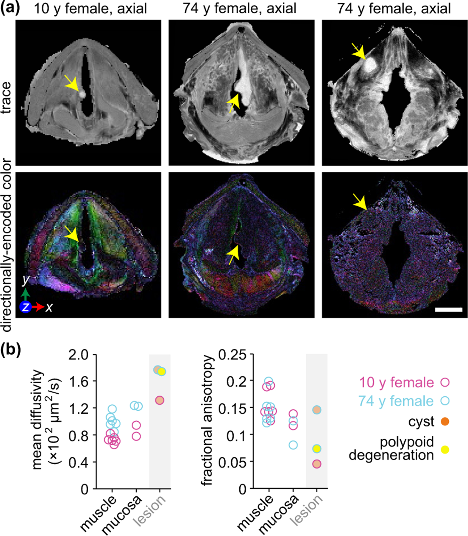
FIGURE 7.

DTI of mucus retention cysts and polypoid degeneration. (a) Trace and directionally encoded colour maps of a infraglottic mucus rentention cyst in a 10-year-old female, polypoid degeneration in a 74-year-old female and supraglottic mucus retention cyst in the same 74-year-old female. The trace maps show the diffusivity of water within the specimens. The directionally encoded colour maps are weighted by anisotropy and show the primary orientation of tissue fibres: green, anterior-posterior; red, medial-lateral; blue, cranial-caudal. Note: The 74-year-old specimen had the lateral thyroid cartilage laminae resected prior to imaging. (b) Mean diffusivity and fractional anisotropy of the three lesions compared to skeletal muscle and mucosa in the same larynges. The muscle and mucosal regions are those shown in Figure 2b. Measurements were obtained via manual tracing of each region of interest in the relevant DTI images from each full larynx scan. DTI, diffusion tensor imaging; y, year. Scale bar, 5 mm
