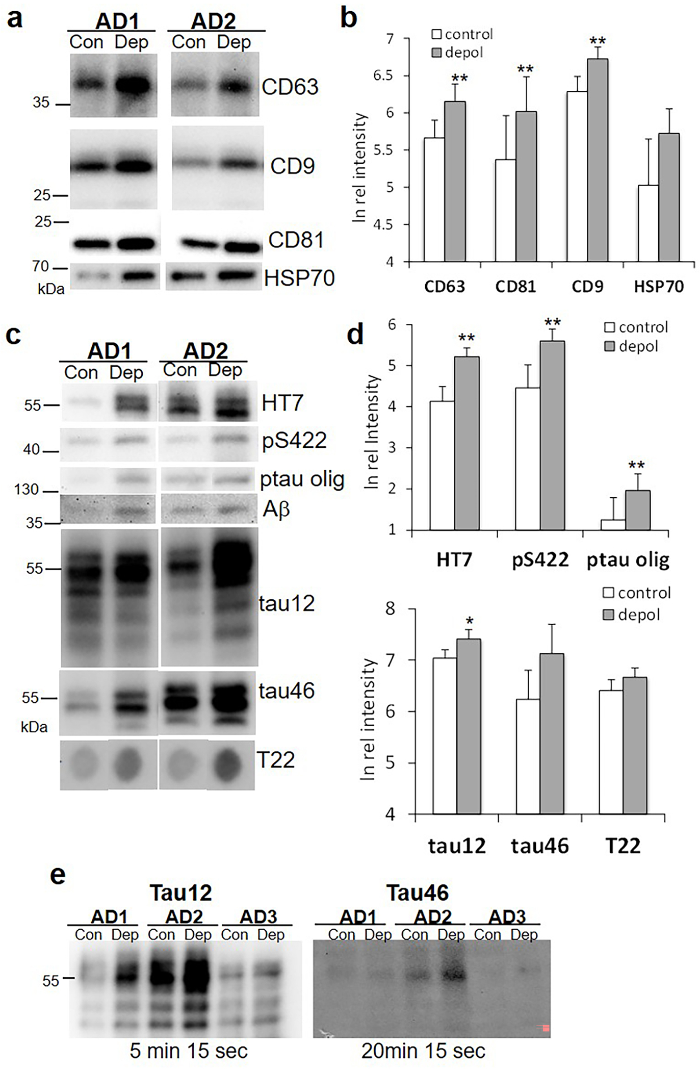
Figure 2. Western SDS PAGE analysis of synaptosome release supernatants.

(a) Representative immunoblots demonstrate labeling for markers of extracellular vesicles: levels of the tetraspanins CD63, CD9, CD81, and heat shock protein 70 (HSP70) are compared in control (con) and depolarized (dep) samples. (b) Aggregate data for (a); n=7, p<0.01. (c) Representative immunoblots for tau peptides (see Table 1 for antibodies); the ptau oligomer was immunolabeled with PS422. For T22 image is a dot blot. (d) Aggregate data for (c); n=6–13, *p<0.05, **p<0.01. (e) Immunoblots with tau12 (detects C-terminal truncated; intact N-terminus), and tau46 (detects N-terminal truncated; intact C-terminus) that include exposure times below blot, showing faint tau46 despite a four-fold longer exposure time. Blots shown are representative of 5 separate experiments with 3–7 cases/blot for each of the two antibodies.
