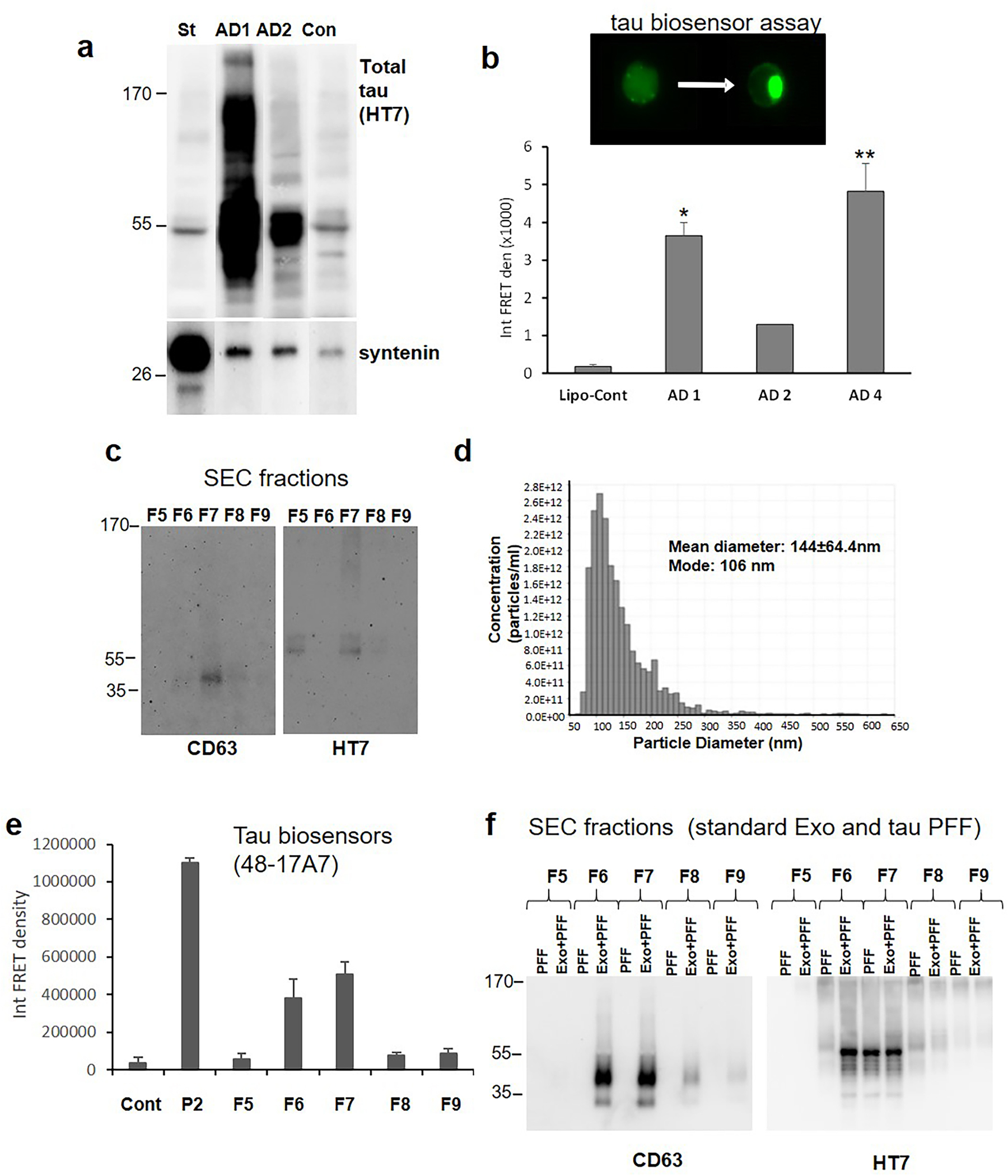
Figure 3. AD cortical synapses release exosomal tau with seeding activity.

(a) Large (~200mg) P2 samples from two AD and one control (Con) cases were depolarized and exosomes from each case were purified by simultaneous immunoprecipitation (IP) with antibodies to CD63, CD9, and CD81 (pan exosome isolation). IP samples were immunolabeled with the HT7 antibody against tau and the exosome marker syntenin; the left lane control (St) shows labeling with commercial standard human plasma exosomes. (b) To determine seeding activity, release supernatants from three P2 samples were concentrated and loaded to HEK293T Tau RD P301S FRET biosensor cells (tau biosensor). Aggregate data (mean± SEM) is shown for the three AD cases along with lipofectamine control (lipo-cont), all in duplicate; *p<0.05, **p<0.01, students t test for independent samples. (c) Representative Western SDS PAGE analysis of SEC fractions shows EV/exosome signal with antibody to tetraspanin CD63 and the total tau antibody HT7. (d) Representative Tunable Resistive Pulse Sensing (TRPS) analysis shows the size of particles in F7 fraction consistent with exosomes. (e) HEK293 tau biosensor assay for SEC fractions; integrated FRET Density, Int FRET den, Cont is lipofectamine control, P2 is crude synaptosome positive control. Error bars represent mean±SEM, p<0.05. (f) Western SDS PAGE of SEC fractions with added standard exosomes (Exo) plus commercial tau fibrils (PFF) alternating with added PFF alone.
