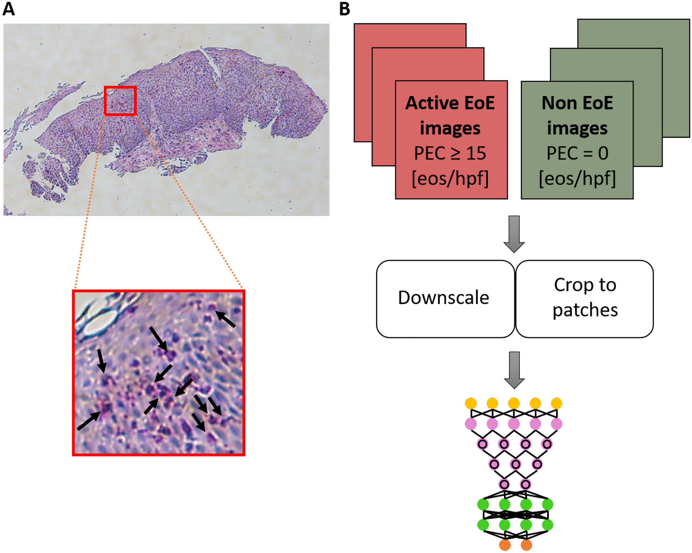
FIGURE 1.

(A) Example of a full-size hematoxylin and eosin (H&E)-stained esophageal biopsy slide from a patient with active eosinophilic esophagitis (EoE). The red square marks an example of an area containing eosinophils (bright pink cells with purple nuclei; several examples are indicated by black arrows in the inset). (B) Schematics of the platform. Images (magnification 80×) of research slides (from one esophageal research biopsy per patient) are labeled as EoE or non-EoE on the basis of a pathologist’s analysis of corresponding clinical slides associated with the same endoscopy during which the research biopsy was obtained. The full-size images are downscaled and/or cropped using various approaches to smaller images that are then used to train a deep convolutional neural network (DCNN). eos, eosinophils; hpf, high-power field; PEC, peak eosinophil count.
