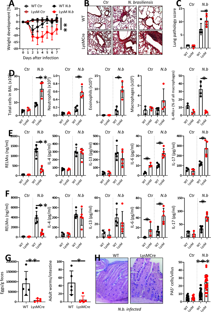
Figure 7. Ptpn2-LysMCre mice are more susceptible to Nippostrongylus brasilensis infection.
7-week-old Ptpn2-LysMCre and their WT littermates were infected with 500 Nippostrongylus brasiliensis L3 stage larvae. A) Weight development post infection, B) representative pictures of lung histology and C) respective scoring, D) numbers of infiltrating cell, neutrophils, eosinophils, and macrophages as well as IL-4Ra expression on macrophages in BAL fluid at day 7. E+F: levels of the indicated cytokines in E) BAL fluid and F) small intestinal homogenate on day 7 post infection. G) Egg and worm count in the small intestine and the feces on day 7. H) Alciane blue-PAS staining and goblet cell counts of small intestinal sections from mice on day 7. N.b. = Nippostrongylous brasiliensis infected. Representative results from one out of two independent experiments with 4–6 mice per group, each. * = p < 0.05, ** = p < 0.01, *** = p < 0.001, Kruskal-Wallis with Dunn’s multiple comparisons test.

