
Figure 1: Dp/E2f1 is required in CySCs to maintain hub cells.
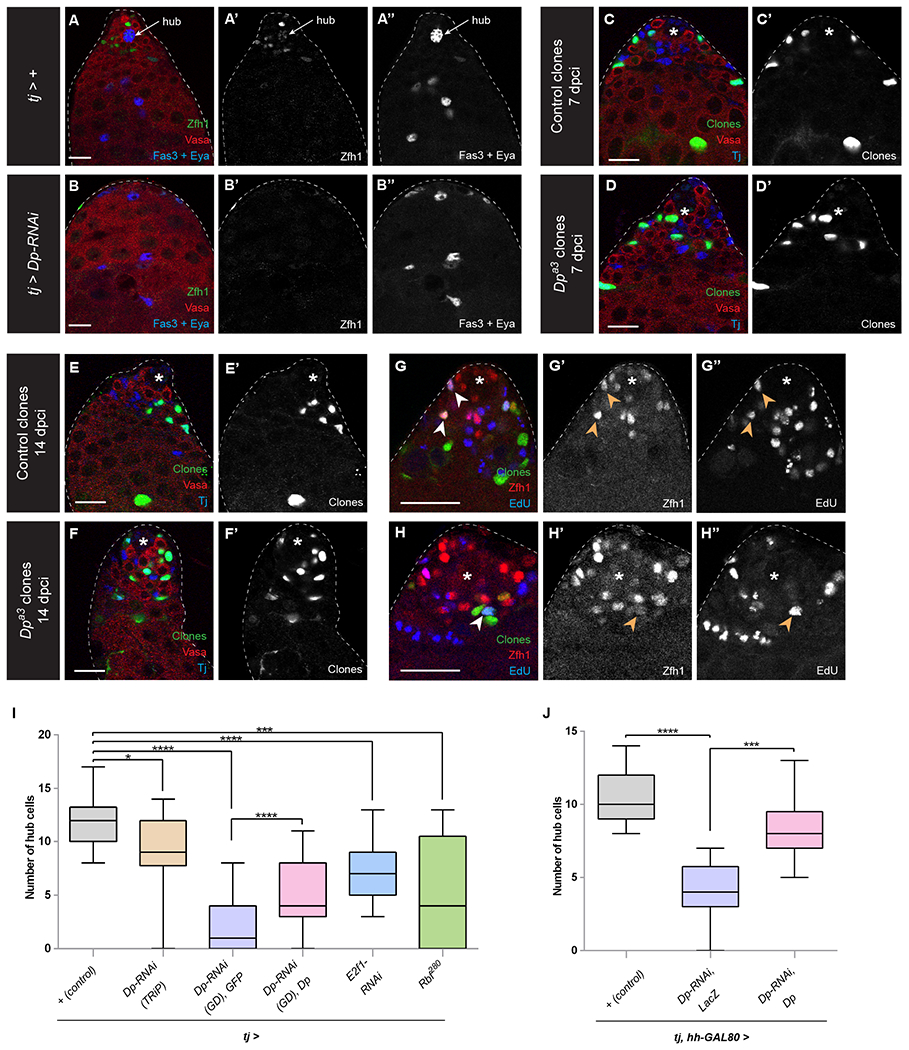
(A, B) A control tj-GAL4 (labeled tj > +) adult testis with hub cells (A, arrow) surrounded by both GSCs and CySCs. A tj > Dp RNAi (B) adult testis lacking CySCs, GSCs and hub cells. Both testes were isolated after 10 days at 29°C to induce maximal GAL4 activity. Zfh1 (green) labels CySCs; Vasa (red) marks the germline; Fas3 (blue) marks the hub cell membranes; Eya (blue) labels the nucleus of differentiating cyst cells.
(C-H) GFP-positive FRT42 control clones (C, E, G) or FRT42 Dpa3 mutant clones (D, F, H). Both types of CySC clones can be recovered at 7 days post clone induction (dpci) (C, D) and 14 dpci (E ,F) and both incorporate EdU (blue G, H), indicating that they can undergo S phase. Clones are marked by GFP (green), Vasa (red, C-F) marks the germline, and Tj (blue, C-F) marks CySCs and early cyst cells. Zfh1 (blue, G, H) marks CySCs.
(I) Graph showing the average number of hub cells after 10 days in 29°C using tj-GAL4 in control (+, gray bar, n=18), Dp-RNAi (brown and purple bars, n=14 and n=31, respectively), Dp depletion plus exogenous Dp (pink bar, n=15), E2f1 depletion (blue bar, n=17), overexpression of Rbf280 (green bar, n=12).
(I, J) Graphs showing the average number of hub cells after 10 days in 29°C using tj-GAL4 and hh-GAL80, which inhibits GAL4 activity in the hub, limiting expression of UAS-dependent constructs to CySCs in control (+, gray bar, n=27), Dp-RNAi (purple bar, n=8), Dp depletion plus exogenous Dp (pink bar, n=33).
An asterisk marks the hub.
Error bars represent the data range. **** P < 0.0001; *** P < 0.001; * P < 0.05 as assessed by Student’s t-test.
See also Tables S1 and S2, Figures S1 and S2.
Scale bar = 20 μM.