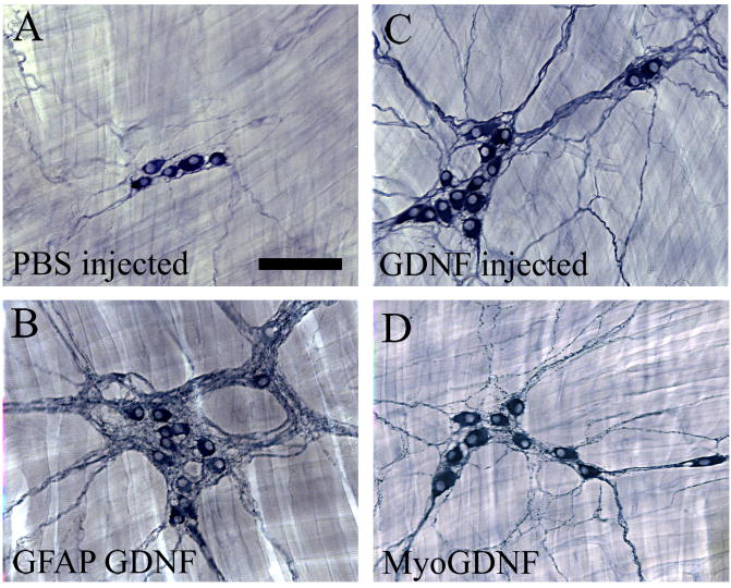
Figure 5. Esophageal innervation is influenced by increased GDNF.
(A) NADPH-d myenteric neurons in WT esophagus or from PBS injected mice are typically found in small clusters with a few small neuronal fibers between ganglia. (B) GFAP-Gdnf mice had more esophageal neurons and a dramatic accumulation of neuronal fibers near enteric ganglia. GDNF injected mice (C) and Myo-Gdnf (D) had more abundant neurons, as well as a higher density of neuronal fibers. Scale bar (100 μm).

