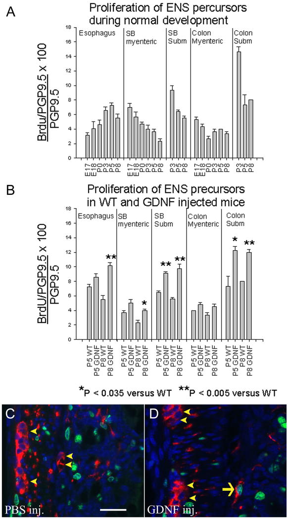
Figure 6. Because ENS precursors have regional and temporal differences in proliferation rates that change during development, increased GDNF leads to increased precursor proliferation in subsets of cells that have not yet exited the cell cycle.
(A) The percentage of PGP9.5 expressing cells that labeled with BrdU was determined in different regions of the bowel at E17, E18, P0, P3, P5 and P8. (B) Proliferation rates for PGP9.5 expressing enteric neurons were also determined in mice injected with GDNF daily from P0 to P5 or P0 to P8. (C, D) PGP9.5/BrdU double label immunohistochemistry in mice injected with PBS (C) or GDNF (D) from P0 to P5. Mice were analyzed at P5 three hours after BrdU injection. Arrow indicates a PGP9.5/BrdU double positive cell. Arrowheads indicate PGP9.5+ but BrdU negative cells. N = 3 mice at each age. Error bars represent ± SEM. *P < 0.035 versus WT; **P < 0.005 versus WT. Scale bar (25μm).

