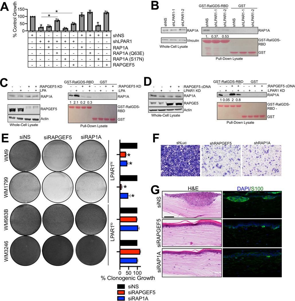
Figure 4.
LPA regulates LPAR1 signaling through a RAPGEF5-RAP1A-axis. (A) MTT assay was performed in WM9 LPAR1 knockdown cells as well as in LPAR1 knockdown cells with the expression of RAP1A (Q63E, constitutively active mutant), RAP1A (S17N, inactive mutant), overexpression of RAP1A (wt) or RAPGEF5. n=3; ANOVA with post-hoc Holm-Sidak’s multiple comparisons test were applied. * Holm-Sidak’s multiple comparisons adjusted p<0.05; error bars indicate SD. (B) GST-tagged RalGDS-RBD was conjugated on glutathione Sepharose 4B and incubated with WM9 cells infected with shLuc, shLPAR1–1 or shLPAR1–2. (C-D) GSTtagged RalGDS-RBD was conjugated on glutathione Sepharose 4B and incubated with WM9 cells with the addition of LPA followed by the knockdown of RAPGEF5 (C) or the overexpression of RAPGEF5 followed by the knockdown of LPAR1 (D). (E) Melanoma cell lines were transfected with siNS, siRAPGEF5 or siRAP1A and grown for 3 weeks in long-term colony formation assays. Cells were subsequently stained with crystal violet and imaged, n=3. ANOVA with post-hoc Holm-Sidak’s multiple comparisons test were applied. * Holm-Sidak’s multiple comparisons adjusted p<0.05; error bars indicate SD. (F) Transwell invasion assay (with matrigel coating) was performed in WM9 cell lines infected with shNS, shRAPGEF5 or shRAP1A. (G) WM9 cells transfected with siNS, siRAPGEF5, or siRAP1A were used to make skin reconstructs. S100, green; DAPI, blue. Scale bar for H&E staining, 50 μM. Scale bar for immunofluorescence staining, 20 μM.

