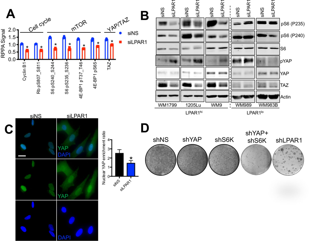
Figure 5.
mTOR and YAP are downstream effectors of the LPAR1-axis. (A) WM9 cells were transfected with siNS or siLPAR1 for 48 hr. Protein lysate was analyzed by RPPA. Shown are the proteins most significantly altered. (B) A panel of LPAR1lo and LPAR1hi melanoma cell lines were transfected with siNS or siLPAR1 for 48 hr. Protein lysate was immunoblotted to validate findings in (A). (C) Immunostaining of YAP in WM9 cells transfected with siNS or siLPAR1 using an anti-YAP antibody (green); nuclei were stained with DAPI (blue). The ratio of YAP localized to the nucleus was quantified and shown to the right. (D) WM9 cells expressing shNS, shYAP, shS6K, shYAP+shS6K, or shLPAR1 were grown for 3 weeks in long-term colony formation assays and subsequently stained with crystal violet. An unpaired two-tailed t-test was used for all studies.

